
Getting to Know CBCT: What You Need to Know
CBCT (Cone Beam Computed Tomography) is a specialized type of X-ray that creates detailed 3D images of your teeth, jaw, and surrounding structures. This guide is perfect for patients who’ve been recommended for a CBCT scan, dental professionals wanting to refresh their knowledge, and anyone curious about this imaging technology.
We’ll break down how CBCT technology actually works and what makes it different from regular X-rays. You’ll also learn about safety measures and radiation exposure levels, plus get a clear picture of what happens during your actual scan appointment. By the end, you’ll feel confident and informed about the entire CBCT process.
Understanding CBCT Technology and How It Works
Definition and Core Principles of Cone Beam Computed Tomography
Cone beam computed tomography represents a specialized imaging technique that captures detailed three-dimensional images of anatomical structures using a cone-shaped X-ray beam. Unlike traditional medical imaging that relies on flat detector systems, CBCT employs a cone-shaped radiation source that rotates around the patient in a single 360-degree sweep, collecting hundreds of planar projection images during this rotation.
The technology works by reconstructing these multiple two-dimensional projections into comprehensive 3D datasets through sophisticated mathematical algorithms. The cone beam configuration allows for simultaneous capture of entire anatomical regions, making the scanning process both efficient and thorough. This approach generates volumetric data that can be manipulated and viewed from virtually any angle, providing medical professionals with unprecedented diagnostic capabilities.
The core principle relies on the geometric relationship between the X-ray source, the patient, and the detector panel. As the imaging system rotates, each projection captures slightly different perspectives of the internal structures. Advanced reconstruction software then processes these images using filtered back-projection or iterative reconstruction techniques to create detailed cross-sectional views, similar to slicing through the anatomy layer by layer.
Key Differences Between CBCT and Traditional CT Scans
CBCT systems operate fundamentally differently from conventional multi-slice CT scanners in several critical ways. Traditional CT machines use a fan-shaped X-ray beam that captures thin slices sequentially, requiring the patient table to move continuously through the scanner gantry. This helical or axial approach demands longer scan times and often results in higher radiation exposure.
| Feature | CBCT | Traditional CT |
|---|---|---|
| X-ray beam shape | Cone-shaped | Fan-shaped |
| Rotation cycles | Single 360° sweep | Multiple rotations |
| Scan time | 10-40 seconds | 1-10 minutes |
| Patient positioning | Seated or standing options | Lying down only |
| Image resolution | Higher spatial resolution | Better soft tissue contrast |
| Radiation dose | Significantly lower | Higher dose levels |
The cone beam approach captures entire anatomical volumes in one rotation, dramatically reducing scan times and patient movement artifacts. CBCT systems also offer greater flexibility in patient positioning, with many units allowing upright scanning that better represents natural anatomical relationships under gravitational loading.
Cost considerations also differ substantially. CBCT systems typically require lower initial investment and reduced facility infrastructure compared to traditional CT scanners, making advanced imaging more accessible to smaller practices and specialized clinics.
Advanced Imaging Capabilities That Benefit Patient Diagnosis
CBCT technology delivers exceptional spatial resolution, often achieving sub-millimeter detail that surpasses traditional CT imaging capabilities. This enhanced resolution proves particularly valuable when examining complex anatomical structures with intricate details, such as temporal bone anatomy, dental roots, or small joint spaces.
The isotropic voxel capability of CBCT means that image resolution remains consistent in all three spatial dimensions, allowing for accurate measurements and assessments regardless of viewing orientation. This uniformity enables precise treatment planning and outcome evaluation across different clinical specialties.
Multi-planar reconstruction capabilities allow clinicians to examine structures in sagittal, coronal, axial, and oblique planes simultaneously. This comprehensive visualization helps identify pathology that might be missed in single-plane imaging and provides better understanding of spatial relationships between different anatomical structures.
Advanced post-processing tools enable various specialized viewing modes, including maximum intensity projections, volume rendering, and curved planar reconstructions. These visualization techniques help highlight specific pathological processes and improve diagnostic confidence by presenting information in clinically relevant formats.
Real-Time 3D Visualization Advantages for Medical Professionals
The ability to manipulate and examine 3D datasets in real-time transforms the diagnostic process for healthcare providers. Interactive visualization allows immediate exploration of anatomical relationships without waiting for additional imaging studies or reconstruction processing.
Measurement tools integrated into CBCT viewing software provide accurate dimensional analysis directly on the 3D images. Clinicians can measure distances, angles, areas, and volumes with precision that supports evidence-based treatment planning and progress monitoring.
Collaborative features enable multiple specialists to review cases simultaneously, even from different locations, fostering interdisciplinary communication and treatment coordination. The ability to share specific views, annotations, and measurements streamlines consultation processes and improves patient care coordination.
Dynamic viewing capabilities allow medical professionals to “fly through” anatomical structures, providing perspective similar to endoscopic visualization without invasive procedures. This virtual navigation enhances understanding of complex anatomy and helps identify optimal surgical approaches or treatment pathways.
Patient Safety and Radiation Exposure Considerations
What to Expect During Your CBCT Scan Experience
Simple Preparation Steps and Pre-Scan Requirements
Getting ready for a CBCT scan is surprisingly straightforward. You won’t need to fast or take any special medications beforehand. Just wear comfortable clothing and avoid metal jewelry around your head and neck area – earrings, necklaces, and hairpins should be removed since metal can interfere with image quality.
If you wear dentures or have removable dental appliances, you might be asked to take them out depending on the scan area. Contact lenses are fine to keep in, but glasses will need to come off. Your dental team will provide a secure place to store your belongings during the procedure.
Women should inform their dentist if there’s any possibility of pregnancy, as radiation exposure needs careful consideration during this time. Bring a list of current medications if you’re taking anything that affects bone density or healing, as this information helps with treatment planning.
Quick and Comfortable Scanning Process Walkthrough
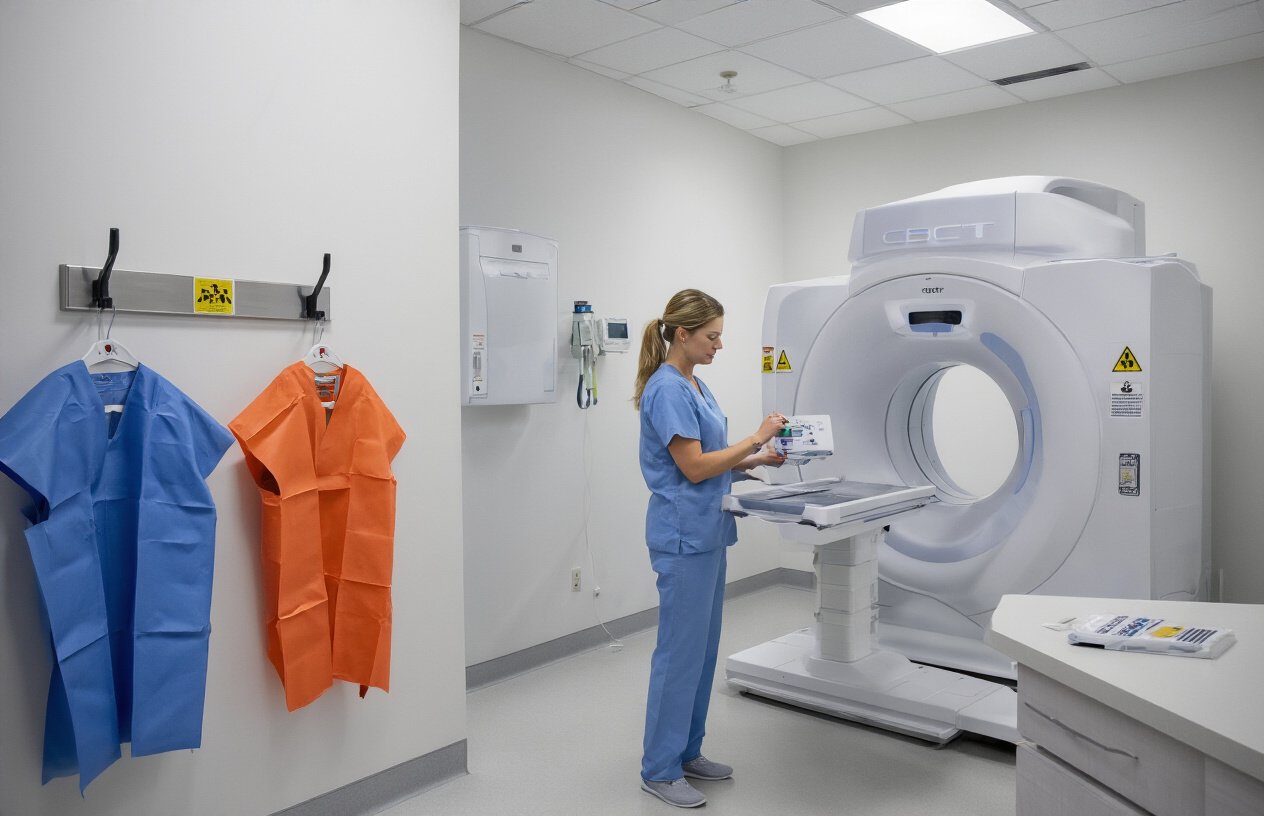
The actual CBCT scan takes just 10-40 seconds of X-ray exposure time, though the entire appointment usually lasts about 15-20 minutes. You’ll be positioned either standing, sitting, or lying down depending on your dental office’s equipment setup.
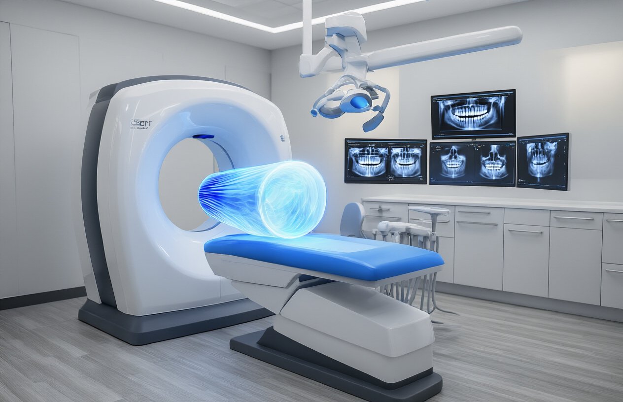
The machine looks like a large donut that rotates around your head. You’ll need to stay perfectly still during the scan – even small movements can blur the images. A chin rest and head stabilizer help keep you in the right position. Some patients find it helpful to focus on breathing slowly through their nose during the brief scanning period.
The technician operates the machine from a separate room but can see and communicate with you through a window. You’ll hear some whirring and clicking sounds as the machine rotates – this is completely normal. Most people find the experience much more comfortable than they expected, especially compared to traditional medical CT scans.
Immediate Image Availability and Consultation Scheduling
One of the biggest advantages of CBCT technology is how quickly your images are ready. Unlike traditional X-rays that might need developing time, your 3D images appear on the computer screen within minutes of completing the scan.
Your dentist can immediately review the detailed images with you, pointing out specific areas of concern and explaining treatment recommendations right there in the office. This real-time consultation means you can ask questions while everything is fresh in your mind and start discussing treatment options during the same visit.
Many practices can schedule follow-up appointments or coordinate with specialists before you leave, streamlining your care process. If you need to share images with other healthcare providers, digital files can be easily transferred or burned to a disc for you to take along.
CBCT technology has transformed how dental professionals diagnose and treat patients by providing detailed 3D images that reveal what traditional X-rays simply can’t show. The cone beam approach delivers precise imaging while keeping radiation exposure at reasonable levels, making it a smart choice for complex dental cases. When you understand the safety protocols and know what happens during the scanning process, you can feel confident about this advanced diagnostic tool.
Ready to experience the benefits of CBCT imaging? Talk to your dentist about whether this technology could help with your specific dental needs. The clearer picture it provides often leads to better treatment outcomes and can save you time and discomfort in the long run.