
CBCT imaging has transformed how oral surgeons approach impacted third molar cases, offering detailed 3D views that traditional X-rays simply can’t match. This technology is particularly valuable for dentists, oral surgeons, and dental specialists who need to assess complex impaction cases where nerve damage or other complications pose significant risks.
This guide is designed for:
- Oral surgeons planning third molar extractions
- General dentists evaluating referral cases
- Dental professionals seeking to understand advanced imaging benefits
While panoramic radiographs remain the standard first step, CBCT becomes essential when signs suggest close contact between the tooth and mandibular canal. The technology helps identify patients at higher risk for nerve injury and guides surgical approach decisions.
We’ll explore three key areas:
Understanding CBCT Technology in Third Molar Assessment – How cone beam imaging provides superior visualization of root-to-nerve relationships compared to traditional 2D radiographs, and when the upgrade from panoramic imaging makes clinical sense.
Critical Risk Assessment Benefits – The specific ways CBCT helps identify potential complications like inferior alveolar nerve injury, adjacent tooth root resorption, and bone loss patterns that aren’t visible on standard radiographs.
Enhanced Treatment Planning and Cost Considerations – How 3D imaging changes surgical approaches, improves patient safety outcomes, and whether the additional cost provides measurable clinical value for both routine and high-risk cases.
Understanding CBCT Technology in Third Molar Assessment
How CBCT Provides Superior 3D Imaging Over Traditional Methods
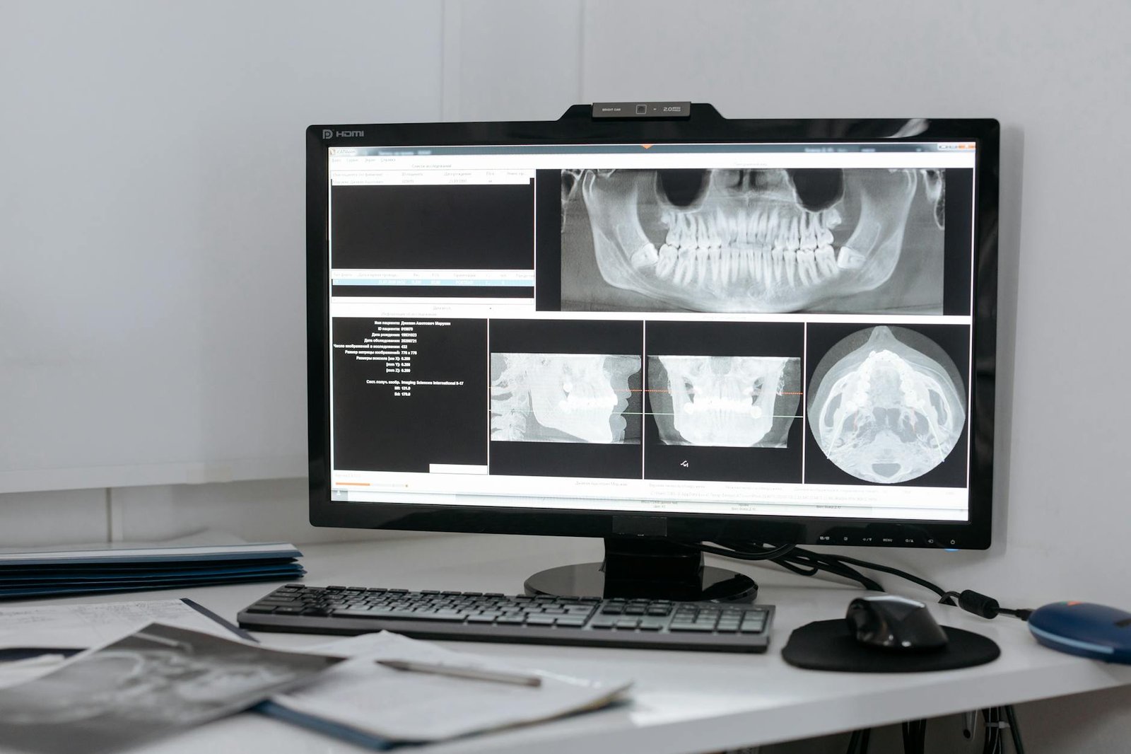
Cone Beam Computed Tomography fundamentally transforms the diagnostic landscape for impacted third molar evaluation through its three-dimensional imaging capabilities. Unlike conventional two-dimensional radiographic methods, CBCT offers comprehensive volumetric data that eliminates the inherent limitations of traditional imaging techniques. The superior information provided by CBCT’s 3D images represents a paradigm shift in how clinicians assess lower third molar surgical cases.
Traditional radiographic methods, including panoramic radiographs and periapical films, present anatomical structures in a compressed, two-dimensional format that often obscures critical spatial relationships. This dimensional limitation frequently leads to diagnostic uncertainty, particularly when evaluating the proximity of impacted third molars to vital anatomical structures such as the mandibular canal. The overlapping of anatomical features in conventional radiography can mask important details that are crucial for surgical planning and risk assessment.
CBCT technology addresses these fundamental limitations by providing detailed cross-sectional views in multiple planes—axial, coronal, and sagittal—along with the ability to generate three-dimensional reconstructions. This comprehensive imaging approach allows clinicians to visualize the exact position of impacted third molars in relation to surrounding anatomical structures without the distortion and superimposition inherent in traditional two-dimensional radiography.
The volumetric nature of CBCT imaging enables precise measurement of distances between the impacted tooth and critical structures such as the inferior alveolar nerve canal. This capability is particularly valuable when assessing the buccal-lingual position of the tooth, which cannot be accurately determined from conventional panoramic radiographs. The three-dimensional perspective provided by CBCT allows for accurate assessment of root morphology, including the presence of dilacerated or curved roots that may complicate surgical extraction.
Furthermore, CBCT imaging provides superior visualization of bone density and trabecular patterns around impacted third molars. This enhanced tissue characterization capability enables more accurate assessment of the surgical difficulty and helps predict potential complications during extraction procedures. The ability to examine the impacted tooth from multiple angles and in various planes significantly enhances diagnostic confidence compared to traditional imaging methods.
The cross-sectional imaging capability of CBCT also reveals important anatomical variations that may not be apparent on conventional radiographs. These variations include accessory canals, bifid inferior alveolar nerves, and unusual root configurations that could significantly impact surgical planning and patient safety. The comprehensive view provided by CBCT ensures that such anatomical variations are identified preoperatively, allowing for appropriate modification of surgical techniques.
When CBCT Becomes Necessary Beyond Standard Panoramic Imaging
The decision to utilize CBCT imaging for impacted third molar evaluation extends beyond routine cases and becomes particularly crucial when specific clinical and radiographic indicators suggest increased surgical complexity or risk. While panoramic radiography remains the initial imaging modality of choice for most third molar assessments, certain situations warrant the enhanced diagnostic capabilities that only CBCT can provide.
CBCT becomes especially indicated when an overprojection is observed between the third molar and the mandibular canal on panoramic radiographs. This radiographic finding suggests potential proximity between the impacted tooth and the inferior alveolar nerve, creating a situation where precise three-dimensional localization becomes critical for safe surgical planning. The overprojection phenomenon occurs when the two-dimensional nature of panoramic imaging creates apparent overlap between structures that may or may not be in actual contact in three-dimensional space.
Specific radiographic signs observed on panoramic images serve as triggers for CBCT evaluation. These signs include darkening of the root where it crosses the mandibular canal, deflection or narrowing of the canal, interruption of the cortical outline of the canal, and bifid appearance of the canal around the root apex. When any of these signs are present, they suggest close contact between the impacted third molar and the mandibular canal, making CBCT imaging essential for accurate risk assessment.
The clinical significance of these radiographic indicators cannot be overstated, as they represent warning signs of potential nerve injury during surgical extraction. The proximity suggested by these signs on two-dimensional imaging requires three-dimensional confirmation to determine the actual spatial relationship between the tooth and nerve structure. CBCT provides this confirmation by clearly delineating the exact position of the mandibular canal in relation to the impacted tooth roots.
Beyond nerve-related considerations, CBCT becomes necessary when conventional imaging fails to provide adequate visualization of root morphology or when complex anatomical relationships are suspected. Cases involving deeply impacted teeth with unclear root patterns, teeth in close proximity to the maxillary sinus, or situations where previous surgical attempts have been unsuccessful often require the enhanced visualization that CBCT provides.
The decision to proceed with CBCT imaging also becomes warranted when treatment planning requires precise measurements for surgical access or when alternative treatment approaches are being considered. For instance, when coronectomy is being contemplated as an alternative to complete extraction, CBCT provides the detailed anatomical information necessary to evaluate the feasibility of this conservative approach.
Additionally, CBCT imaging becomes essential in cases where patient-specific factors increase the risk of complications. These factors include advanced patient age, presence of medical conditions that affect bone healing, or previous history of complications with oral surgical procedures. In such cases, the comprehensive information provided by CBCT enables more accurate risk stratification and informed treatment planning.
The timing of CBCT imaging also becomes crucial in complex cases where initial treatment planning based on conventional radiography proves inadequate. When intraoperative findings differ significantly from preoperative expectations based on two-dimensional imaging, CBCT can provide valuable information for modifying surgical approach or planning subsequent procedures.
Technical Capabilities That Enhance Diagnostic Accuracy
The technical specifications and capabilities of CBCT systems directly contribute to enhanced diagnostic accuracy in third molar assessment through multiple advanced features that surpass traditional imaging modalities. These technical capabilities work synergistically to provide clinicians with unprecedented visualization of anatomical structures and pathological conditions associated with impacted third molars.
The high spatial resolution achievable with CBCT technology represents one of its most significant technical advantages. Modern CBCT systems can achieve voxel sizes as small as 0.1 millimeters, providing exceptional detail resolution that enables visualization of fine anatomical structures such as the lamina dura around tooth roots and the cortical boundaries of the mandibular canal. This level of detail is particularly important when assessing the integrity of anatomical boundaries that may be compromised by impacted teeth.
The contrast resolution capabilities of CBCT systems allow for differentiation between various tissue types and pathological conditions. This capability is essential for identifying pericoronitis, cystic lesions, or other pathological conditions associated with impacted third molars. The ability to distinguish between different soft tissue densities and identify areas of bone sclerosis or rarefaction provides valuable diagnostic information that influences treatment planning decisions.
However, studies examining the efficacy of radiographic methods, including CBCT, acknowledge that much of the supporting evidence operates at lower levels within established evidence hierarchies. Specifically, research evaluating CBCT’s technical capabilities and diagnostic accuracy often falls within Levels 1-3 of the six-tiered evidence model, indicating that while the technology shows promise, continued research is needed to establish definitive clinical guidelines.
The multiplanar reconstruction capabilities of CBCT systems enable clinicians to examine anatomical structures from virtually unlimited perspectives. This flexibility in viewing angles is particularly valuable when assessing complex spatial relationships between impacted teeth and surrounding structures. The ability to generate curved planar reconstructions along the path of the mandibular canal provides unique visualization opportunities that are impossible with conventional imaging techniques.
Advanced image processing algorithms incorporated into modern CBCT systems enhance diagnostic accuracy through noise reduction, artifact minimization, and contrast optimization. These processing capabilities improve image quality and reduce the likelihood of diagnostic errors caused by technical limitations. The ability to adjust image parameters post-acquisition allows for optimization of visualization for specific diagnostic tasks.
The volume rendering capabilities of CBCT systems enable three-dimensional visualization of anatomical structures, providing an intuitive understanding of spatial relationships that is particularly valuable for surgical planning. These rendering techniques can highlight specific structures of interest while maintaining visualization of surrounding anatomy, creating customized views that enhance diagnostic interpretation.
Furthermore, the measurement tools available in CBCT software provide precise quantitative assessment capabilities that enhance diagnostic accuracy. These tools enable accurate measurement of distances, angles, and volumes, providing objective data that supplements qualitative visual assessment. The ability to measure the minimum distance between impacted tooth roots and the mandibular canal provides specific quantitative information for risk assessment.
The technical capability for iterative image reconstruction in CBCT systems allows for optimization of image quality based on specific diagnostic requirements. This flexibility enables clinicians to balance radiation dose with image quality requirements, ensuring that diagnostic accuracy is maintained while minimizing patient radiation exposure.
Cross-sectional imaging capabilities provided by CBCT technology eliminate the superimposition artifacts that commonly affect conventional radiographic interpretation. This elimination of overlapping structures significantly reduces diagnostic uncertainty and enhances the accuracy of anatomical assessment. The ability to examine structures in isolation from overlapping anatomy provides clearer visualization of pathological conditions and anatomical variations.
The archival and communication capabilities of digital CBCT systems enhance diagnostic accuracy through improved image storage, retrieval, and sharing. High-quality digital images can be easily transmitted for consultation purposes, enabling collaboration between specialists and improving overall diagnostic accuracy through collective expertise. The ability to manipulate and enhance digital images post-acquisition provides additional opportunities for optimizing diagnostic interpretation.
Critical Risk Assessment Benefits of CBCT

Identifying Proximity Between Tooth Roots and Inferior Alveolar Canal
CBCT technology provides unparalleled visualization of the spatial relationship between impacted third molar roots and the inferior alveolar canal, a critical anatomical structure that houses the inferior alveolar nerve. This three-dimensional imaging capability represents a fundamental advancement in pre-surgical assessment, offering clinicians the precise anatomical information necessary to evaluate potential complications before they occur.
The inferior alveolar canal runs through the mandible, carrying the inferior alveolar nerve, artery, and vein. Traditional two-dimensional radiographic techniques often fail to accurately depict the exact proximity of impacted third molar roots to this vital structure. CBCT addresses this limitation by providing detailed cross-sectional images that clearly delineate the canal’s pathway and its relationship to the tooth roots in question.
When examining impacted third molars, the proximity assessment becomes crucial for determining surgical approach and risk stratification. CBCT clearly identifies the proximity of the tooth root to the inferior alveolar canal, enabling clinicians to measure exact distances and understand the three-dimensional spatial relationships that would otherwise remain obscured in conventional imaging. This precise visualization allows for accurate determination of whether the canal runs buccally, lingually, or directly through the root structure.
The superior imaging quality of CBCT reveals subtle anatomical variations that significantly impact surgical planning. Some patients present with canals that bifurcate or demonstrate unusual pathways around impacted teeth. These anatomical variations, which are virtually impossible to detect with traditional radiography, become clearly visible with CBCT imaging. Understanding these variations is essential for preventing inadvertent nerve damage during extraction procedures.
Furthermore, CBCT imaging allows clinicians to assess the cortication of the inferior alveolar canal. A well-corticated canal indicates intact bone surrounding the nerve structure, while loss of cortication suggests closer proximity or potential contact with the tooth roots. This information directly influences surgical technique selection and helps determine whether specialized procedures, such as coronectomy, might be more appropriate than complete tooth removal.
The ability to precisely measure distances between root apices and the canal wall provides quantitative data that supports clinical decision-making. Research has established threshold distances that correlate with increased risk of nerve injury, and CBCT enables accurate measurement of these critical distances. This objective assessment tool transforms subjective clinical judgment into evidence-based risk stratification.
Preventing Inferior Alveolar Nerve Injury Complications
Now that we have covered the anatomical visualization capabilities of CBCT, it becomes evident how this technology directly contributes to preventing one of the most serious complications associated with third molar extraction: inferior alveolar nerve injury. CBCT aids in risk assessment prior to surgery, contributing to improved safety and treatment outcomes and potentially preventing complications like inferior alveolar nerve injury.
Inferior alveolar nerve injury can result in temporary or permanent numbness, tingling, or altered sensation in the lower lip, chin, and teeth on the affected side. These complications can significantly impact patient quality of life, affecting functions such as eating, drinking, speaking, and kissing. The psychological impact of such complications cannot be understated, as patients may experience anxiety, depression, and social withdrawal due to altered facial sensation.
The comprehensive risk assessment enabled by CBCT allows clinicians to identify high-risk cases before surgery begins. When CBCT imaging reveals close proximity between tooth roots and the inferior alveolar canal, several surgical modifications can be implemented to minimize nerve injury risk. These may include altered surgical approaches, coronectomy procedures where only the crown is removed while leaving roots in place, or staged surgical techniques that allow for gradual tooth movement away from the nerve.
CBCT imaging also facilitates better informed consent discussions with patients. By clearly visualizing the anatomical relationships and quantifying risks, clinicians can provide patients with accurate information about potential complications. This enhanced communication allows patients to make truly informed decisions about their treatment, understanding both the benefits and risks associated with different surgical approaches.
The prevention of nerve injury through CBCT-guided planning extends beyond immediate surgical considerations. Long-term follow-up studies have demonstrated that patients who undergo CBCT-guided third molar extractions experience significantly lower rates of permanent nerve damage compared to those treated based on conventional radiographic assessment alone. This evidence underscores the clinical value of investing in advanced imaging technology for complex third molar cases.
Additionally, CBCT enables the identification of patients who might benefit from referral to oral and maxillofacial surgeons with specialized expertise in complex extractions. General dentists can use CBCT findings to recognize cases that exceed their comfort level or require specialized surgical techniques, ensuring that patients receive the most appropriate level of care for their specific anatomical situation.
Evaluating Adjacent Second Molar Root Resorption Risks
With this in mind, next, we’ll examine how CBCT technology addresses another critical concern in impacted third molar management: the assessment of root resorption risks to adjacent second molars. CBCT allows for the assessment of potential root resorption of the adjacent second molar, providing crucial information that influences treatment timing and approach decisions.
Root resorption of the adjacent second molar represents a progressive condition where the impacted third molar causes dissolution of the root structure of the neighboring tooth. This process typically occurs slowly and may remain asymptomatic until significant damage has occurred. Traditional two-dimensional radiography often fails to detect early stages of root resorption, particularly when the resorption occurs on the buccal or lingual surfaces of the root that are not clearly visible in standard radiographic projections.
CBCT imaging reveals root resorption in its earliest stages by providing complete three-dimensional visualization of the second molar root structure. This comprehensive assessment allows clinicians to detect subtle changes in root morphology that indicate the beginning of resorptive processes. Early detection is crucial because it enables intervention before irreversible damage occurs to the second molar.
The detailed imaging provided by CBCT allows for precise assessment of resorption extent and location. Clinicians can determine whether resorption affects the cervical, middle, or apical third of the root, information that directly impacts the prognosis of the affected second molar. Surface resorption involving only the outer root surface may be compatible with long-term tooth retention, while deep resorption extending into the pulp space typically necessitates endodontic treatment or tooth extraction.
CBCT also enables monitoring of root resorption progression over time. By comparing sequential CBCT images, clinicians can assess whether resorption is actively progressing or has stabilized. This longitudinal assessment capability is particularly valuable for patients who are not immediate candidates for third molar extraction due to medical or social factors, allowing for continued monitoring until treatment becomes feasible.
The three-dimensional nature of CBCT imaging helps distinguish true root resorption from radiographic artifacts or anatomical variations that might mimic resorptive changes in conventional radiography. This differential diagnostic capability prevents unnecessary treatments based on false-positive findings while ensuring that genuine resorption is not overlooked.
Furthermore, CBCT assessment of root resorption influences surgical planning for third molar extraction. When significant resorption is present, surgical approach may need modification to minimize additional trauma to the compromised second molar. In some cases, the presence of extensive resorption may contraindicate certain surgical techniques or require simultaneous treatment of both the third and second molars.
Assessing Bone Loss at Distal Aspects
Previously, we’ve discussed the soft tissue and root structure assessments enabled by CBCT technology. Now, we must address another critical evaluation capability: the assessment of bone loss patterns around impacted third molars. CBCT helps in assessing bone loss at the distal aspects of the adjacent second molar as a consequence of the third molar, providing essential information for comprehensive treatment planning and long-term prognosis determination.
Bone loss at the distal aspect of the second molar represents a common consequence of impacted third molar presence, resulting from chronic inflammation, bacterial accumulation, and mechanical pressure effects. This bone loss can compromise the long-term stability and health of the second molar, potentially leading to periodontal complications, increased mobility, and eventual tooth loss if left unaddressed.
CBCT imaging provides detailed visualization of bone architecture around both the impacted third molar and the adjacent second molar. The three-dimensional perspective allows clinicians to assess bone loss patterns with unprecedented accuracy, revealing both the extent and morphology of defects that would be impossible to fully appreciate with conventional radiographic techniques.
The assessment of distal bone loss involves evaluation of several critical parameters. CBCT enables measurement of bone loss depth from the cemento-enamel junction of the second molar to the most apical extent of the defect. This quantitative assessment provides objective data for treatment planning and helps establish baseline measurements for monitoring progression or healing following intervention.
Additionally, CBCT reveals the three-dimensional configuration of bone defects, distinguishing between horizontal bone loss, vertical defects, and combination patterns. This morphological information directly influences treatment approach, as different defect types respond differently to various regenerative procedures and surgical techniques.
The width and circumferential extent of bone loss around the second molar distal surface can be precisely measured using CBCT imaging. This information helps determine whether bone loss is localized to a small area or extends around a significant portion of the root circumference, affecting treatment prognosis and complexity.
CBCT also enables assessment of bone quality and density in areas affected by chronic inflammation. The imaging can reveal areas of sclerotic bone formation, which may indicate chronic infection or inflammatory processes that require specific treatment approaches. Conversely, areas of decreased bone density may suggest active infection or rapid bone destruction requiring immediate intervention.
The relationship between bone loss patterns and the position of the impacted third molar becomes clearly visible with CBCT imaging. This information helps clinicians understand the mechanism of bone destruction and predict potential for spontaneous healing following third molar removal. In cases where significant bone loss has occurred, CBCT assessment helps determine whether additional regenerative procedures will be necessary to restore adequate bone support for the second molar.
Furthermore, CBCT assessment of bone loss extends to evaluation of the overall alveolar process integrity. The imaging can reveal whether bone loss is limited to the immediate area around the third molar or has extended to compromise broader areas of the mandible or maxilla. This comprehensive assessment is crucial for understanding the full scope of treatment required and predicting long-term outcomes.
The detailed bone assessment provided by CBCT also helps identify cases where bone grafting or regenerative procedures may be beneficial following third molar extraction. By understanding the extent and configuration of bone defects prior to surgery, clinicians can prepare appropriate grafting materials and plan simultaneous regenerative procedures to optimize healing outcomes for the adjacent second molar.
Enhanced Treatment Planning and Decision-Making
Cost-Effectiveness and Clinical Impact Considerations
Determining When CBCT Changes Treatment Outcomes
Previously, we’ve explored the enhanced treatment planning capabilities of CBCT technology. Now that we have covered the clinical advantages and risk assessment benefits, it’s crucial to understand when CBCT imaging truly makes a difference in patient outcomes and whether the additional cost and radiation exposure can be justified from both clinical and economic perspectives.
The fundamental principle guiding CBCT utilization in impacted third molar evaluation centers on whether the additional imaging will alter treatment decisions or improve patient outcomes. This evidence-based approach requires careful consideration of when conventional two-dimensional radiography proves insufficient and when three-dimensional imaging becomes clinically necessary.
Evidence Levels and Clinical Decision Making
The assessment of CBCT’s clinical impact operates within a six-tiered evidence model that provides a framework for evaluating the true value of diagnostic imaging technologies. Higher-evidence studies, specifically those at Levels 4, 5, and 6 of this model, focus on the most clinically relevant outcomes that extend beyond simple image quality or diagnostic accuracy measures.
Level 4 studies assess the diagnostic impact of radiographic methods, examining whether the additional information provided by CBCT actually changes the diagnostic process or clinical decision-making pathway. These investigations determine if the superior visualization capabilities of CBCT translate into altered treatment recommendations when compared to conventional radiographic approaches.
Level 5 evidence evaluates patient outcomes directly, measuring whether the enhanced diagnostic capabilities of CBCT result in improved clinical results for patients undergoing third molar extraction. This level of evidence examines tangible benefits such as reduced complications, decreased post-operative morbidity, shorter healing times, or improved functional outcomes.
The highest tier, Level 6 evidence, incorporates societal implications and comprehensive cost calculations. These studies examine the broader healthcare economic impact, considering not only the direct costs of CBCT imaging but also the downstream effects on treatment efficiency, complication rates, and overall healthcare resource utilization.
Current Research Landscape and Evidence Gaps
Despite the widespread adoption of CBCT technology in oral and maxillofacial surgery, there are very few high-evidence studies specifically examining the efficacy of CBCT for third molar evaluation. This research gap represents a significant limitation in our understanding of when CBCT truly provides clinical value beyond conventional radiographic methods.
The scarcity of high-level evidence studies indicates a need for further research at higher evidence levels, particularly studies that can demonstrate concrete improvements in patient outcomes and cost-effectiveness. This research deficit means that clinical decisions regarding CBCT utilization often rely on lower-level evidence, such as case series, expert opinions, or studies focused primarily on image quality rather than clinical outcomes.
This evidence gap creates challenges for clinicians attempting to make evidence-based decisions about when to recommend CBCT imaging. Without robust outcome data, practitioners must rely on clinical judgment and extrapolation from available research to determine when the additional information provided by CBCT will translate into meaningful improvements in patient care.
Clinical Scenarios Where CBCT Demonstrates Impact
The principle that CBCT is indicated when it is believed that its use will change the treatment plan or treatment outcome provides a framework for identifying appropriate clinical scenarios. This criterion requires practitioners to assess whether the additional three-dimensional information will alter their therapeutic approach or improve the likelihood of successful outcomes.
Complex anatomical relationships represent one category where CBCT often changes treatment outcomes. When impacted third molars demonstrate close proximity to critical anatomical structures such as the inferior alveolar nerve, maxillary sinus, or adjacent tooth roots, the superior visualization provided by CBCT can significantly alter surgical approach and technique. In these cases, the three-dimensional information may lead to modified extraction techniques, alternative treatment timing, or even decisions to monitor rather than extract.
Cases involving unusual tooth morphology or root configuration present another scenario where CBCT frequently impacts treatment planning. When conventional radiographs suggest complex root anatomy, dilacerated roots, or unusual crown-to-root relationships, CBCT can provide crucial information that alters surgical planning and instrumentation choices.
Suspected pathology associated with impacted third molars represents a critical application where CBCT often changes treatment outcomes. When conventional radiographs suggest the presence of cysts, tumors, or other pathological processes, CBCT can provide essential information about the extent and nature of the lesion, fundamentally altering treatment planning and potentially involving additional specialists.
Cost-Effectiveness Analysis Framework
Determining the cost-effectiveness of CBCT in impacted third molar evaluation requires a comprehensive analysis that extends beyond the immediate imaging costs. The economic evaluation must consider multiple factors, including the direct costs of CBCT acquisition and interpretation, the potential for altered treatment planning, and the downstream effects on clinical outcomes and healthcare resource utilization.
Direct imaging costs include not only the expense of CBCT acquisition but also the professional fees for interpretation and the time required for image review and treatment planning. These costs must be weighed against the potential benefits of enhanced diagnostic information and improved treatment planning capabilities.
Indirect cost considerations encompass the potential for CBCT to prevent complications that would require additional treatment and healthcare resources. When CBCT helps avoid inferior alveolar nerve injuries, sinus perforations, or damage to adjacent teeth, the prevention of these complications can result in significant cost savings through reduced need for additional procedures, specialist referrals, or long-term management of complications.
The cost-effectiveness calculation must also consider the impact on surgical efficiency and outcomes. If CBCT imaging leads to more precise surgical planning, reduced operative time, or improved first-attempt success rates, these benefits can translate into economic advantages through improved practice efficiency and reduced need for revision procedures.
Societal and Healthcare System Implications
The broader societal implications of CBCT utilization in third molar evaluation extend beyond individual patient costs to encompass healthcare system efficiency and resource allocation. When CBCT imaging prevents complications or improves treatment outcomes, the benefits extend to reduced healthcare system burden and improved resource utilization.
Population-level considerations include the appropriate allocation of advanced imaging resources and ensuring that CBCT technology is utilized in cases where it provides the greatest clinical benefit. This requires careful consideration of imaging protocols and selection criteria that maximize the clinical impact while minimizing unnecessary radiation exposure and healthcare costs.
The societal perspective also encompasses the long-term implications of diagnostic imaging decisions. When CBCT prevents nerve injuries that could result in permanent sensory deficits, the societal benefits include not only the direct healthcare cost savings but also the preservation of quality of life and functional capacity for affected individuals.
Clinical Decision-Making Framework
With this in mind, next, we’ll see how practitioners can develop a systematic approach to determining when CBCT is likely to change treatment outcomes. This decision-making framework should incorporate assessment of clinical complexity, evaluation of conventional radiographic adequacy, and consideration of the potential for altered treatment planning based on additional three-dimensional information.
The framework begins with thorough evaluation of conventional radiographic findings and identification of specific clinical questions that remain unanswered. When panoramic or intraoral radiographs provide adequate information for safe and effective treatment planning, additional imaging may not be justified. However, when conventional imaging leaves critical questions about anatomical relationships, pathology, or surgical complexity unanswered, CBCT may be indicated.
Risk stratification plays a crucial role in this decision-making process. Patients presenting with high-risk scenarios, such as close proximity to neurovascular structures, suspected pathology, or complex anatomical relationships, are more likely to benefit from CBCT imaging. Conversely, routine extractions with clear anatomical relationships and low complexity may not require advanced imaging.
The decision-making framework must also incorporate patient-specific factors, including age, medical history, and individual risk tolerance. Younger patients with developing root structures may benefit from CBCT evaluation to optimize timing of intervention, while older patients with completed root development and clear anatomical relationships may not require additional imaging.
Future Research Directions and Evidence Development
The recognized need for further research at higher evidence levels presents opportunities for advancing our understanding of CBCT’s clinical impact in third molar evaluation. Future studies should focus on patient outcome measures, cost-effectiveness analyses, and long-term follow-up to determine the true value of three-dimensional imaging in this clinical application.
Randomized controlled trials comparing CBCT-guided treatment planning to conventional radiographic approaches represent the gold standard for generating high-level evidence. These studies should examine not only immediate surgical outcomes but also long-term complications, patient satisfaction, and economic implications.
Prospective cohort studies tracking patients over extended periods can provide valuable insights into the long-term benefits of CBCT-guided treatment planning. These investigations can assess whether the additional diagnostic information translates into sustained improvements in patient outcomes and quality of life.
Economic analyses incorporating comprehensive cost calculations and societal perspectives are essential for determining the true value proposition of CBCT in third molar evaluation. These studies should consider both direct and indirect costs, including the economic impact of prevented complications and improved treatment efficiency.
The development of evidence-based guidelines for CBCT utilization requires synthesis of research findings across multiple study types and evidence levels. As higher-quality evidence becomes available, these guidelines can provide more definitive recommendations for when CBCT imaging is most likely to improve patient outcomes and provide economic value.
CBCT technology represents a significant advancement in mandibular third molar assessment, offering three-dimensional visualization that enhances risk evaluation and treatment planning capabilities. When traditional panoramic imaging reveals potential proximity between the third molar and mandibular canal, CBCT provides critical information about nerve positioning, potential root resorption of adjacent teeth, and bone loss patterns that can directly impact surgical outcomes and patient safety.
The evidence suggests that while panoramic radiographs remain sufficient for most third molar evaluations, CBCT should be considered when specific risk indicators are present and when the additional three-dimensional information will meaningfully influence treatment decisions or improve patient outcomes. Dental professionals must carefully weigh the diagnostic benefits against cost considerations, ensuring that CBCT utilization is justified by its potential to enhance surgical planning and reduce complications like inferior alveolar nerve injury. As the technology continues to evolve, establishing clear clinical protocols for CBCT indication will be essential for optimizing both patient care and resource allocation in oral surgery practice.