
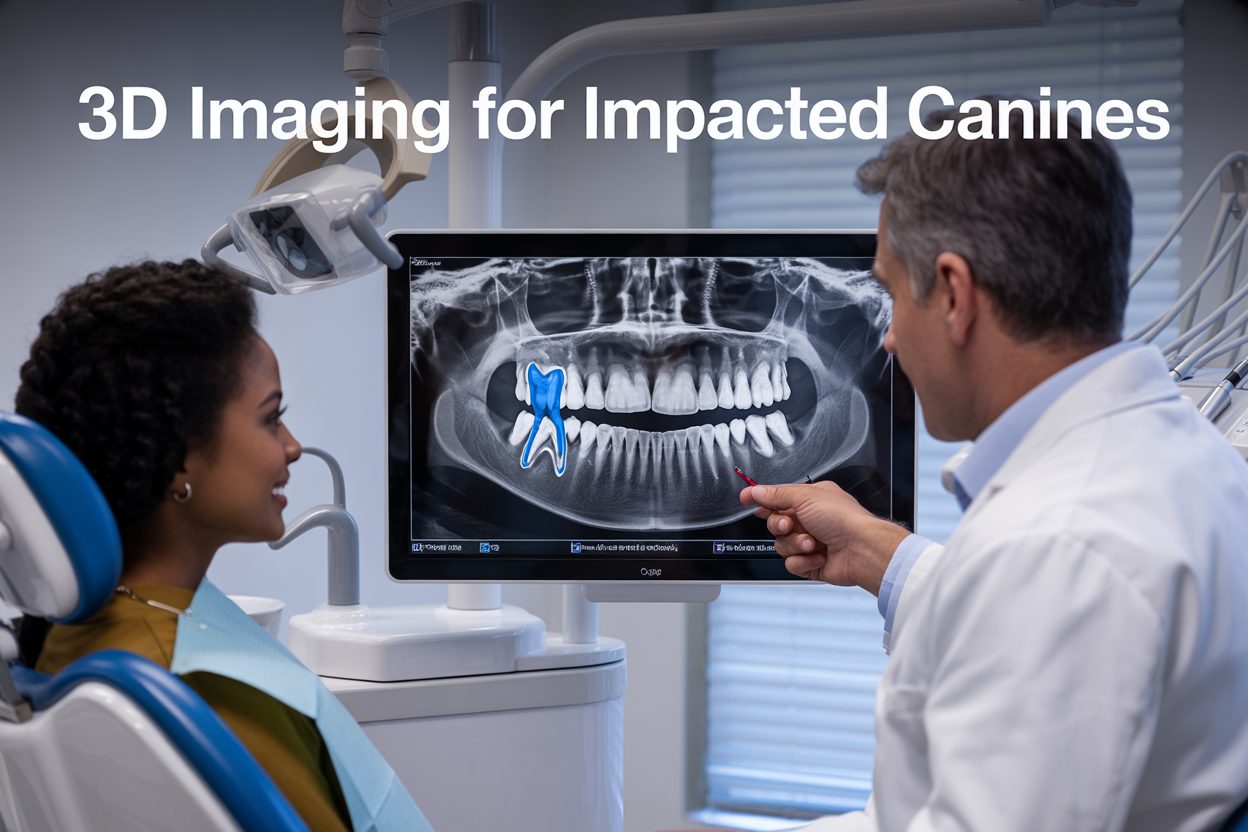
Impacted canines affect up to 3% of the population and present one of the most challenging diagnostic puzzles in dentistry. When these essential teeth fail to erupt properly, precise localization becomes critical for successful treatment outcomes. Traditional X-rays often leave dentists guessing about exact tooth position, root orientation, and potential complications.
This comprehensive guide targets orthodontists, oral surgeons, and general dentists who want to master impacted canine localization using advanced 3D imaging techniques. You’ll discover how CBCT canine impaction analysis revolutionizes diagnosis compared to conventional methods, and learn practical protocols that improve treatment planning accuracy.
We’ll explore the clinical significance of impacted canines and why traditional 2D imaging falls short for complex cases. You’ll also learn step-by-step 3D dental imaging protocols that deliver consistent results, plus advanced analysis techniques that transform raw CBCT data into actionable treatment insights. Finally, we’ll examine real clinical benefits that make orthodontic 3D imaging an essential tool for modern dental practice.
Understanding Impacted Canines and Their Clinical Significance
Traditional 2D Imaging Limitations in Canine Localization
Discover Why Conventional Radiographs Provide Incomplete Information
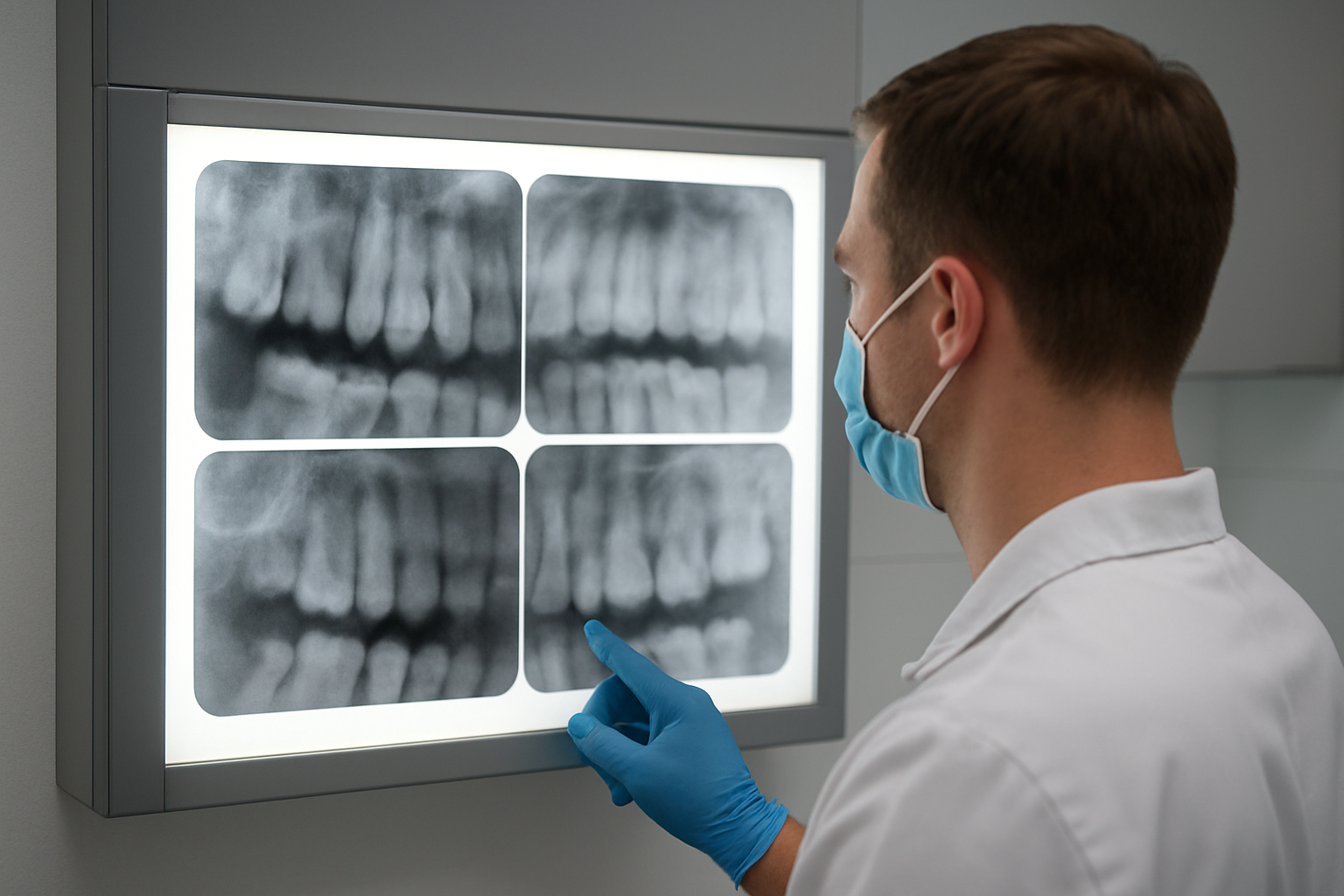
Conventional periapical and panoramic radiographs have been the standard for impacted canine diagnosis for decades, but they present significant challenges when it comes to precise localization. These traditional 2D images compress three-dimensional anatomical structures into a flat representation, causing critical spatial information to disappear completely.
The primary issue lies in the loss of depth perception. When orthodontists examine a standard radiograph, they can see that an impacted canine exists, but determining whether it sits closer to the palatal or buccal surface becomes a guessing game. This uncertainty directly impacts treatment planning decisions and can lead to inappropriate surgical approaches.
Traditional radiographs also struggle with magnification inconsistencies. The distance between the X-ray source, patient, and film creates varying degrees of magnification across different areas of the image. What appears to be a tooth positioned at a certain distance from adjacent structures might actually be significantly closer or farther away in reality.
Root resorption assessment becomes particularly challenging with 2D imaging. While radiographs might show some evidence of lateral incisor root damage, the full extent and exact location of resorption often remain hidden until surgical exposure reveals the true clinical situation.
Learn About Overlapping Structures That Obscure Accurate Positioning
Anatomical superimposition represents one of the most frustrating aspects of conventional canine localization techniques. The maxillary region contains numerous overlapping structures that create confusing shadows and false impressions on 2D radiographs.
The zygomatic process frequently overlaps with impacted canines, creating dense radiopaque areas that mask important anatomical details. This overlap makes it nearly impossible to accurately assess the crown position relative to adjacent tooth roots or determine the angulation of the impacted tooth.
Nasal cavity borders and maxillary sinus walls add another layer of complexity. These structures create intersecting radiopaque and radiolucent lines that can easily be mistaken for tooth outlines or root boundaries. Clinicians often find themselves squinting at radiographs, trying to differentiate between actual tooth structures and anatomical noise.
Adjacent tooth roots compound the visualization problem. When multiple teeth are present in the same vertical plane, their roots create a web of overlapping shadows. Distinguishing the impacted canine’s root from neighboring lateral incisors, first premolars, or central incisors becomes a puzzle that even experienced practitioners find challenging.
The anterior nasal spine frequently projects over the impacted canine region, creating additional confusion. This bony prominence can mask crown positioning and make accurate measurements nearly impossible to obtain from conventional radiographs.
Understand Measurement Errors in Traditional Imaging Methods
Geometric distortion inherent in 2D radiographic systems introduces substantial measurement errors that compromise treatment planning accuracy. The cone-beam projection geometry causes objects positioned at different depths to appear at varying sizes, making distance calculations unreliable.
Angulation errors during radiograph acquisition significantly impact measurement precision. Even slight deviations in horizontal or vertical tube positioning can alter the apparent position of an impacted canine by several millimeters. These seemingly small discrepancies translate into major clinical consequences during surgical procedures.
The parallax effect compounds measurement difficulties when using multiple radiographic projections. While the Clark’s rule (tube shift technique) helps determine labial versus palatal positioning, it provides only general directional information rather than precise spatial coordinates needed for optimal impacted canine treatment planning.
Linear measurements between anatomical landmarks suffer from projection errors that vary depending on the patient’s positioning and the X-ray beam orientation. Distance calculations between the impacted canine crown and adjacent root surfaces often prove inaccurate when verified during actual surgical procedures.
Traditional imaging methods also struggle with reproducibility. Slight variations in patient head positioning between appointments result in different measurements from the same impacted tooth, making it difficult to track changes over time or compare pre-treatment and post-treatment positions accurately.
Revolutionary 3D Imaging Technologies for Precise Localization
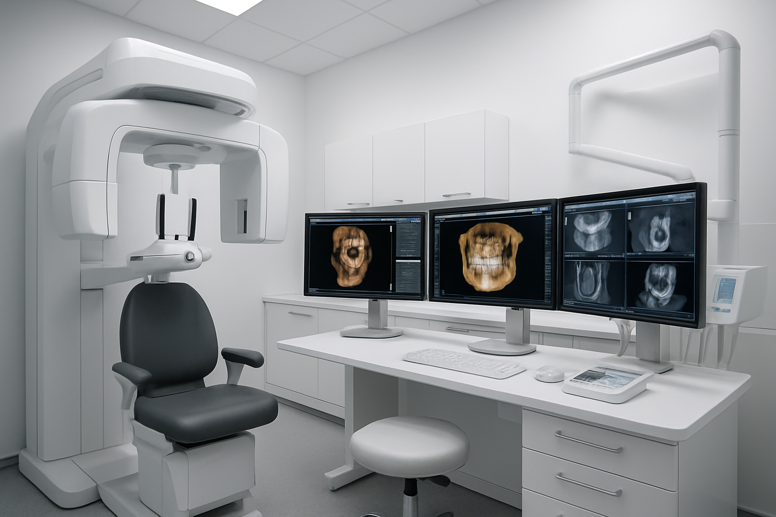
Explore CBCT Scanning Capabilities for Comprehensive Visualization
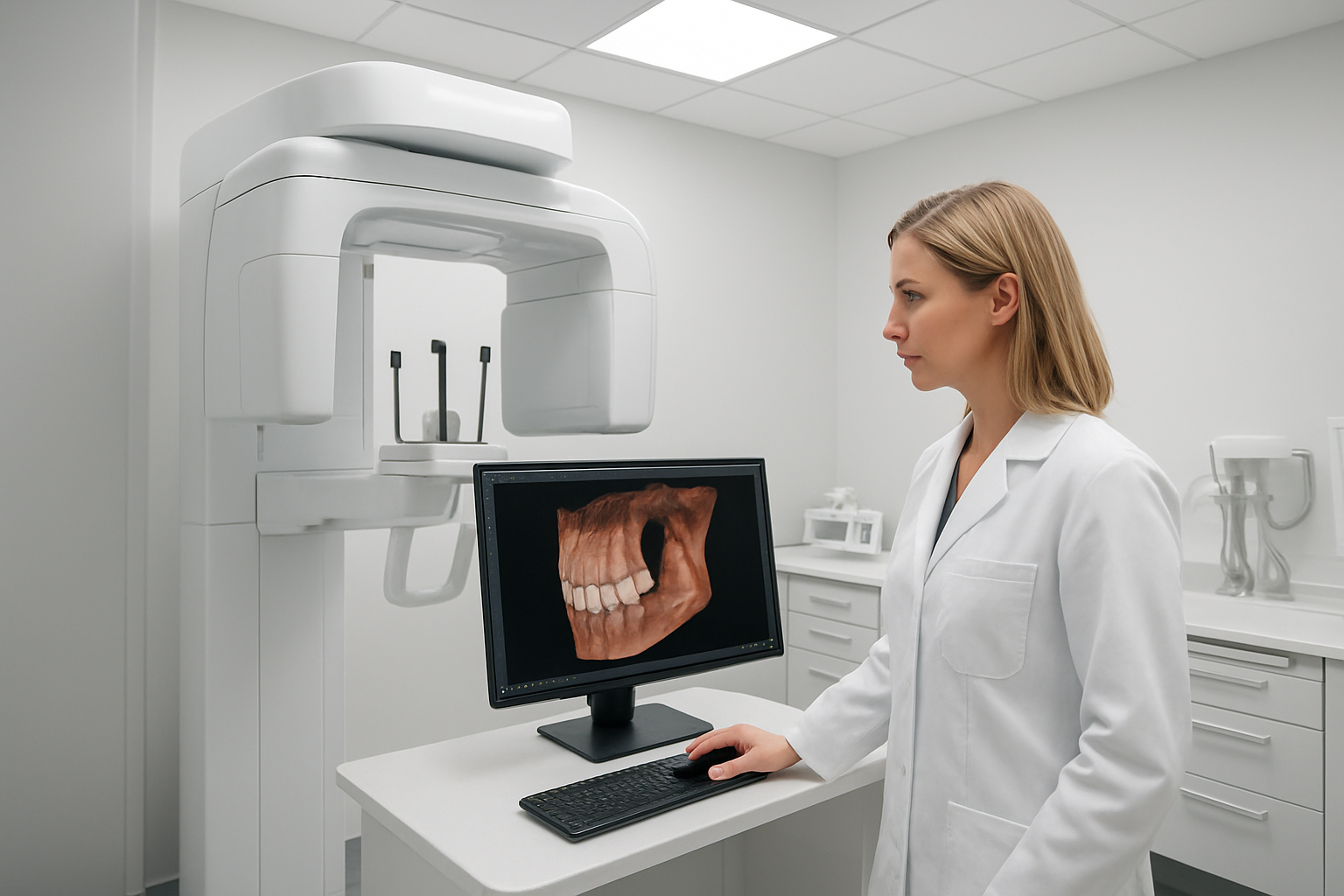
Cone Beam Computed Tomography represents a game-changing advancement in impacted canine localization and 3D dental imaging. Unlike traditional 2D radiographs that compress three-dimensional structures into flat images, CBCT canine impaction studies provide clinicians with unprecedented detail about tooth position, root morphology, and surrounding anatomical structures.
Modern CBCT scanners capture images in seconds while patients remain seated upright, making the process comfortable and efficient. The technology generates high-resolution volumetric data that reveals the precise spatial relationship between impacted canines and adjacent teeth, nerve pathways, and vital structures. This comprehensive visualization enables orthodontists and oral surgeons to assess root resorption risks, plan surgical approaches, and predict treatment timelines with remarkable accuracy.
CBCT imaging excels in revealing critical details that directly impact treatment decisions. Clinicians can visualize the exact angle of impaction, measure distances to neighboring tooth roots, and identify potential complications before surgery begins. The isotropic voxel resolution ensures consistent image quality across all viewing planes, allowing for precise measurements and reliable impacted canine diagnosis.
Discover CT Imaging Advantages for Complex Cases
Medical-grade CT scanning offers superior capabilities for challenging impacted canine localization scenarios where maximum detail and precision are essential. While CBCT dominates routine orthodontic applications, multi-slice CT provides enhanced soft tissue contrast and exceptional bone detail that proves invaluable in complex cases involving extensive pathology, multiple impactions, or anatomical anomalies.
CT imaging delivers submillimeter accuracy in measuring distances and angles, which becomes critical when planning surgical approaches near sensitive structures like the nasopalatine canal or maxillary sinus. The technology’s superior contrast resolution helps differentiate between various tissue types, making it easier to identify cystic lesions, inflammatory changes, or other pathological conditions that might complicate treatment.
For patients with syndromic conditions or craniofacial anomalies affecting multiple teeth, CT scanning provides the comprehensive anatomical information needed for coordinated interdisciplinary treatment planning. The wide field of view captures the entire craniofacial complex, enabling specialists to assess global relationships and plan treatments that address both functional and aesthetic concerns.
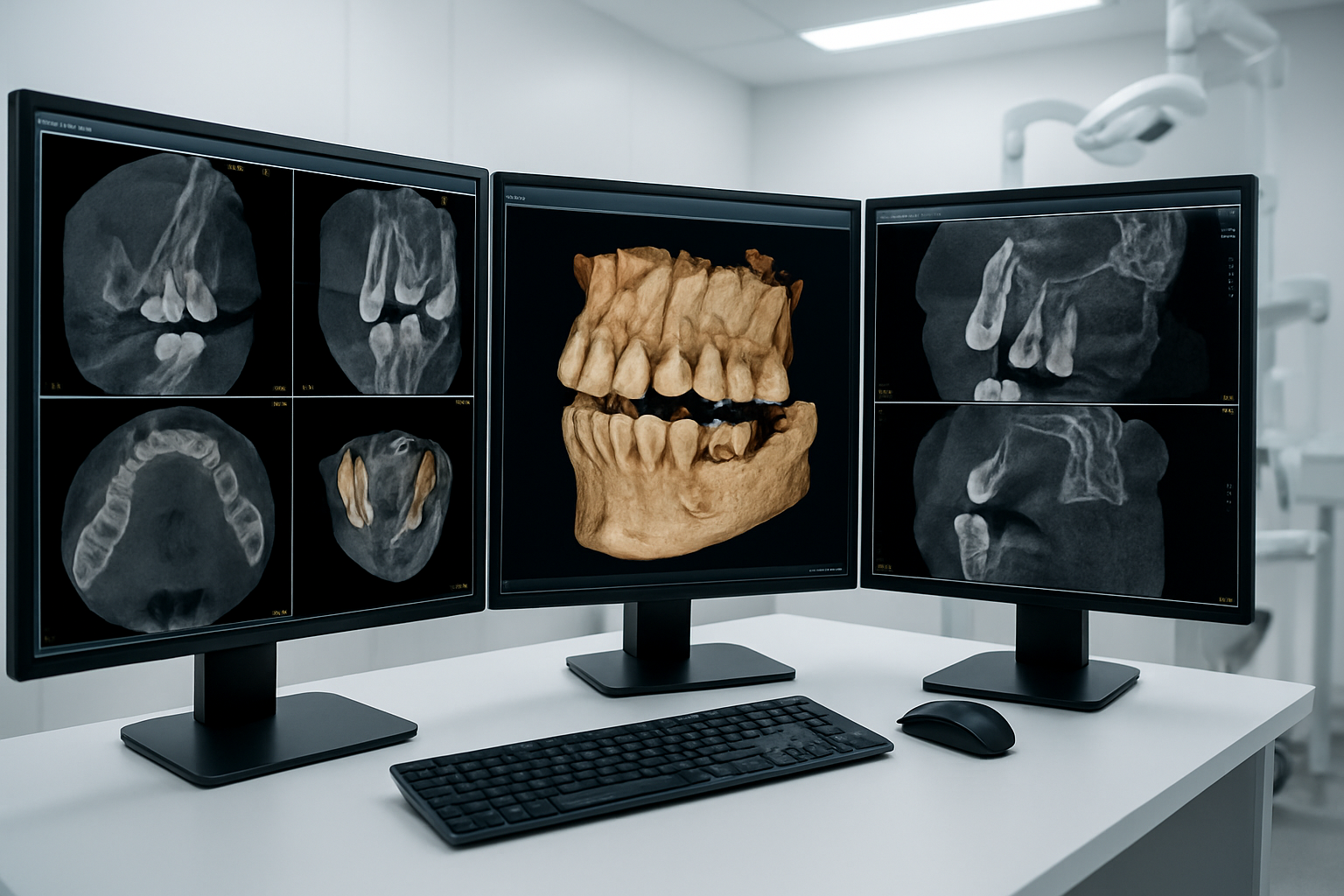
Learn About Advanced Software Tools for 3D Reconstruction
Modern 3D dental imaging relies heavily on sophisticated software platforms that transform raw scan data into actionable clinical information. Leading software solutions offer intuitive interfaces that allow clinicians to manipulate 3D volumes, create precise measurements, and generate detailed treatment plans for impacted canine treatment planning.
These advanced tools provide multiple visualization modes including volume rendering, multiplanar reconstruction, and curved cross-sectional views. Clinicians can virtually dissect anatomical structures, simulate surgical procedures, and create patient-specific guides for optimal treatment outcomes. Many platforms integrate seamlessly with CAD/CAM systems, enabling the fabrication of custom appliances and surgical guides.
Orthodontic 3D imaging software often includes specialized modules for cephalometric analysis, airway assessment, and treatment simulation. These features help practitioners communicate treatment options effectively with patients while ensuring predictable results through detailed pre-treatment planning.
Understand Radiation Dose Considerations in Different Imaging Modalities
Patient safety remains paramount when selecting appropriate impacted canine diagnosis imaging protocols. CBCT technology has revolutionized 3D imaging dentistry by providing detailed anatomical information while maintaining radiation doses significantly lower than traditional medical CT scans.
| Imaging Modality | Typical Effective Dose | Equivalent Background Exposure |
|---|---|---|
| Panoramic X-ray | 14-24 μSv | 2-4 days |
| Small FOV CBCT | 19-65 μSv | 3-11 days |
| Medium FOV CBCT | 87-206 μSv | 15-35 days |
| Large FOV CBCT | 68-1073 μSv | 11-180 days |
| Medical CT Head | 2000 μSv | 11 months |
Optimizing imaging protocols involves balancing diagnostic quality with radiation exposure through careful selection of field of view, voxel size, and scanning parameters. Modern CBCT units incorporate dose reduction technologies like pulsed exposure and automatic exposure control that maintain image quality while minimizing patient exposure. The ALARA principle (As Low As Reasonably Achievable) guides all imaging decisions, ensuring patients receive maximum diagnostic benefit with minimal radiation risk.
Step-by-Step 3D Imaging Protocol for Optimal Results
Advanced Analysis Techniques Using 3D Data
Measure precise distances to adjacent tooth roots
The real game-changer in 3D dental imaging comes when clinicians can measure exact distances between impacted canines and neighboring teeth. Unlike traditional radiographs that compress three-dimensional structures into flat images, CBCT canine impaction analysis provides millimeter-accurate measurements in all spatial planes.
Modern software allows practitioners to draw virtual lines and measure distances directly on the 3D volume. Root proximity measurements become critical when determining if the impacted canine might cause root resorption of adjacent incisors or premolars. Distances less than 2mm typically indicate high risk for root damage during surgical exposure.
The software can automatically calculate the shortest distance between the impacted tooth crown and surrounding root surfaces. This precision helps surgeons plan their approach to minimize trauma to healthy teeth while accessing the impacted canine safely.
Assess proximity to vital anatomical structures
Impacted canine localization requires careful evaluation of nearby critical structures. The maxillary canine’s position relative to the nasal cavity, maxillary sinus, and nasopalatine canal determines surgical risk levels and influences treatment decisions.
3D analysis reveals the exact relationship between the impacted tooth and the nasopalatine canal, which contains important neurovascular structures. When canines are positioned too close to this canal, alternative treatment approaches may be necessary to avoid nerve damage.
For mandibular canines, proximity to the mental foramen and inferior alveolar nerve becomes the primary concern. Advanced software can highlight these structures in different colors, making it easier to visualize safe zones for surgical access.
Determine optimal surgical approach angles
3D imaging dentistry transforms surgical planning by allowing practitioners to simulate different approach angles before making the first incision. The software can calculate the most direct path to the impacted tooth while avoiding damage to surrounding structures.
Surgeons can rotate the 3D image to view the impacted canine from multiple angles, identifying the safest entry point and trajectory. This visualization helps determine whether a palatal, labial, or combined approach would be most effective.
The angle of impaction relative to the adjacent teeth also influences the orthodontic mechanics needed for tooth movement. Severely angulated canines may require different bracket placement or auxiliary springs compared to vertically impacted teeth.
Evaluate bone density around impacted teeth
Bone quality assessment becomes straightforward with impacted canine 3D analysis. The software can measure bone density values in Hounsfield units around the impacted tooth, providing insights into healing potential and surgical difficulty.
Dense cortical bone may require more aggressive surgical techniques or longer healing periods, while areas of low bone density might indicate cystic changes or resorption. This information helps predict surgical complexity and post-operative healing times.
| Bone Density Range | Clinical Interpretation | Surgical Considerations |
|---|---|---|
| >1000 HU | Dense cortical bone | May require piezoelectric surgery |
| 400-1000 HU | Normal trabecular bone | Standard surgical techniques |
| <400 HU | Low density/pathology | Investigate for cysts or resorption |
Create detailed treatment planning maps
The final step in impacted canine treatment planning involves creating comprehensive visual guides that integrate all gathered data. These treatment maps serve as blueprints for the entire interdisciplinary team.
Digital treatment maps can include surgical access routes, orthodontic attachment points, and predicted tooth movement paths. The maps help coordinate care between oral surgeons and orthodontists, reducing treatment time and improving outcomes.
Color-coded risk zones highlight areas requiring special attention during surgery. Green zones indicate safe areas for instrumentation, yellow zones require caution, and red zones represent high-risk areas where vital structures could be damaged.
These detailed planning maps can be shared electronically between specialists and serve as reference guides throughout the treatment process. They also provide excellent documentation for insurance purposes and patient education.
Clinical Benefits That Transform Patient Outcomes
Achieve more predictable surgical procedures
3D imaging dentistry transforms surgical planning by providing unprecedented visualization of impacted canine positions. Surgeons can now examine the exact spatial relationship between impacted teeth and critical anatomical structures like nerve canals, adjacent tooth roots, and sinus cavities before making the first incision.
CBCT canine impaction imaging reveals the precise angulation, depth, and orientation of impacted teeth, eliminating surgical guesswork. Practitioners can determine the optimal surgical approach, whether palatally or labially, and identify the most conservative pathway to expose the crown. This detailed pre-surgical mapping allows orthodontists and oral surgeons to collaborate effectively, creating treatment plans that minimize tissue trauma while maximizing access.
The predictability extends to determining the feasibility of orthodontic traction versus surgical removal. 3D dental imaging clearly shows whether adequate bone support exists for successful orthodontic movement and helps predict the direction and magnitude of forces needed for eruption guidance.
Reduce operative time through better pre-planning
Impacted canine localization using advanced 3D analysis dramatically cuts surgical time by eliminating exploratory procedures. Traditional 2D radiographs often require surgeons to “hunt” for impacted teeth during surgery, extending operative time and increasing patient discomfort.
Comprehensive pre-operative planning with CBCT imaging allows surgical teams to:
- Determine exact entry points for surgical access
- Identify the shortest pathway to the impacted crown
- Anticipate potential complications before they occur
- Pre-select appropriate surgical instruments and techniques
- Coordinate with orthodontic team members for immediate traction placement
Digital treatment planning software enables virtual surgery rehearsals, where practitioners can practice complex procedures before entering the operating room. This preparation translates to smoother, faster procedures with fewer unexpected challenges.
Minimize complications and post-operative discomfort
3D imaging significantly reduces the risk of iatrogenic damage to adjacent structures during impacted canine treatment. Detailed visualization of root proximity prevents accidental damage to neighboring teeth, while clear identification of nerve pathways minimizes the risk of paresthesia or other sensory complications.
Common complications that 3D imaging helps prevent include:
| Complication Type | How 3D Imaging Helps |
|---|---|
| Root resorption | Shows exact proximity to adjacent teeth |
| Nerve damage | Maps inferior alveolar nerve location |
| Sinus perforation | Reveals sinus floor relationship |
| Excessive bone removal | Identifies minimal access pathways |
| Wrong surgical approach | Determines optimal entry angle |
Patients experience less post-operative swelling, pain, and healing time when surgeries are precisely planned and executed using impacted canine diagnosis data from 3D imaging. The ability to create smaller, more targeted surgical sites reduces tissue trauma and accelerates recovery.
Improve long-term treatment success rates
Canine localization techniques using 3D imaging contribute to significantly higher treatment success rates by enabling evidence-based treatment decisions. Orthodontists can accurately predict treatment duration, appliance requirements, and final outcomes before beginning therapy.
Long-term stability improves when impacted canines are guided into their proper positions using biomechanically sound force systems planned with 3D data. The imaging reveals optimal anchor tooth selection, force vectors, and staging sequences that promote healthy periodontal responses and stable final positions.
Orthodontic 3D imaging also helps identify cases where extraction may be more appropriate than attempted eruption, preventing lengthy unsuccessful treatments. This early decision-making protects patients from extended treatment times and potential complications while achieving better overall oral health outcomes.
Treatment success rates for impacted canine management increase from approximately 85% with traditional methods to over 95% when comprehensive 3D treatment planning guides the therapeutic approach.
Getting clear, precise images of impacted canines doesn’t have to be guesswork anymore. 3D imaging has completely changed how we locate these tricky teeth, moving us away from the limitations of traditional 2D X-rays that often left us squinting at unclear shadows. With cone beam CT and other advanced 3D technologies, we can now see exactly where impacted canines sit in relation to neighboring teeth and vital structures, making treatment planning so much more predictable.
The real game-changer is how this technology improves patient care. When you can pinpoint the exact position and angulation of an impacted canine before starting treatment, you’re setting yourself up for success. Patients benefit from shorter treatment times, fewer complications, and better overall outcomes. If you’re still relying solely on 2D imaging for impacted canine cases, it’s time to embrace 3D technology – your patients will thank you for the precision and predictability it brings to their treatment journey.