
Artificial Intelligence in CBCT Interpretation: Present & Future
Artificial intelligence is changing how dental professionals read and analyze CBCT scans, making diagnosis faster and more accurate than ever before. This guide is designed for dentists, oral surgeons, radiologists, and practice managers who want to understand how AI technology can transform their diagnostic workflow and patient care.
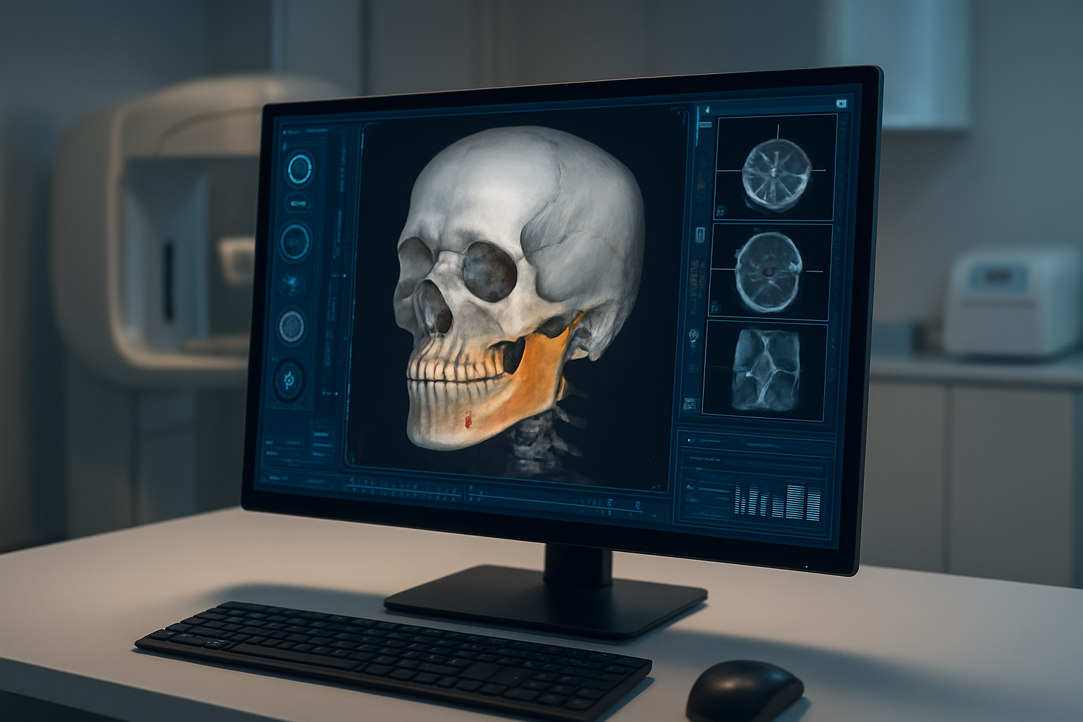
CBCT interpretation has traditionally required extensive training and experience to spot subtle abnormalities and anatomical variations. Now, AI algorithms can highlight potential issues, measure anatomical structures automatically, and even suggest treatment approaches based on scan data.
We’ll explore the current state of AI technology in CBCT analysis and examine the key applications already transforming dental practices today. You’ll also discover the emerging technologies that will shape tomorrow’s CBCT interpretation capabilities and learn about a strategic implementation roadmap to help your practice adopt these powerful tools effectively.
Whether you’re considering your first AI integration or looking to expand existing technology, this overview will help you make informed decisions about incorporating artificial intelligence into your diagnostic process.
Current State of AI Technology in CBCT Analysis
Machine Learning Algorithms Revolutionizing Image Processing
Machine learning has completely transformed how dental professionals approach CBCT image analysis. These sophisticated algorithms can process three-dimensional dental imaging data with remarkable precision, identifying patterns and structures that might escape human detection. The technology builds on decades of research in computer vision and pattern recognition, adapted specifically for dental applications.
Modern CBCT systems generate enormous amounts of data with each scan. A typical CBCT examination produces hundreds of cross-sectional images, each containing millions of voxels representing different tissue densities. Machine learning algorithms excel at managing this data complexity, systematically analyzing every voxel to extract meaningful diagnostic information. The algorithms use mathematical models trained on thousands of previously analyzed CBCT scans to recognize normal anatomical structures and pathological conditions.
The foundation of machine learning in CBCT interpretation rests on supervised learning techniques. These methods require extensive training datasets where expert radiologists have manually annotated anatomical landmarks, pathological findings, and diagnostic conclusions. The algorithms learn to associate specific image patterns with particular diagnoses, gradually building sophisticated decision-making capabilities that mirror expert clinical judgment.
Convolutional neural networks represent the most successful machine learning approach for CBCT analysis. These specialized algorithms excel at processing grid-like data structures, making them perfect for medical imaging applications. CNNs automatically identify relevant features in CBCT images without requiring manual programming of specific detection rules. The networks learn hierarchical representations, starting with simple edge detection and progressing to complex anatomical structure recognition.
Feature extraction algorithms play a crucial role in transforming raw CBCT data into meaningful diagnostic information. These systems identify key characteristics such as bone density variations, root canal morphology, periodontal ligament space widening, and cortical bone integrity. Advanced algorithms can quantify these features with mathematical precision, providing objective measurements that support clinical decision-making.
Texture analysis algorithms have proven particularly valuable for detecting subtle pathological changes in CBCT images. These systems analyze the mathematical relationships between neighboring pixels, identifying patterns that indicate disease processes. For example, the algorithms can detect early signs of bone remodeling around dental implants or identify changes in trabecular bone structure that suggest pathological conditions.
Segmentation algorithms automatically identify and isolate specific anatomical structures within CBCT volumes. These systems can accurately delineate tooth boundaries, distinguish between enamel, dentin, and pulp chambers, and separate individual roots in multi-rooted teeth. The precision of automated segmentation often exceeds manual tracing, providing more consistent and reproducible results across different operators and institutions.
Registration algorithms align CBCT images from different time points, enabling precise comparison of treatment progress or disease development. These systems compensate for patient positioning differences and equipment variations, ensuring accurate longitudinal analysis. The technology proves invaluable for monitoring implant healing, orthodontic tooth movement, and endodontic treatment outcomes.
Preprocessing algorithms enhance CBCT image quality before diagnostic analysis. These systems reduce noise, correct for beam hardening artifacts, and optimize contrast resolution. Machine learning algorithms learn to distinguish between imaging artifacts and genuine pathological findings, preventing false positive diagnoses that could lead to unnecessary treatment.
Classification algorithms categorize anatomical structures and pathological findings according to established diagnostic criteria. These systems can distinguish between different types of cysts, classify fracture patterns, and identify various stages of periodontal disease. The algorithms provide consistent diagnostic terminology and grading systems, improving communication between practitioners and supporting evidence-based treatment planning.
Edge detection algorithms identify boundaries between different tissue types in CBCT images. These systems excel at delineating the periodontal ligament space, detecting root fractures, and outlining pathological lesions. Advanced edge detection methods can identify subtle changes in bone density that indicate early stages of disease progression.
Morphological analysis algorithms quantify the shape and size characteristics of anatomical structures. These systems measure root length, assess alveolar bone height, and evaluate sinus volume for implant planning. The algorithms provide standardized measurements that eliminate inter-operator variability and support objective treatment planning decisions.
Ensemble methods combine multiple machine learning algorithms to improve diagnostic accuracy and reliability. These approaches leverage the strengths of different algorithmic approaches while compensating for individual limitations. Ensemble methods often achieve superior performance compared to single algorithm implementations, providing more robust and consistent diagnostic results.
Transfer learning techniques enable machine learning algorithms trained on large general medical imaging datasets to adapt quickly to specific dental applications. This approach reduces the amount of dental-specific training data required and accelerates algorithm development for specialized diagnostic tasks. Transfer learning has proven particularly valuable for rare pathological conditions where limited training examples are available.
Real-time processing algorithms enable immediate analysis of CBCT images during acquisition. These systems provide instant feedback about image quality and can detect major pathological findings before the patient leaves the imaging facility. Real-time analysis supports point-of-care decision-making and reduces the need for repeat examinations.
Uncertainty quantification algorithms provide confidence measures for diagnostic predictions. These systems indicate when machine learning predictions are reliable and when human expert review is recommended. Uncertainty quantification improves clinical workflow efficiency by prioritizing cases that require immediate attention while allowing routine cases to proceed with automated analysis.
Multi-modal integration algorithms combine CBCT data with other imaging modalities such as intraoral radiographs, clinical photographs, and digital impressions. These systems provide comprehensive diagnostic assessments that consider all available clinical information. Multi-modal approaches often achieve superior diagnostic performance compared to single-modality analysis.
Adaptive learning algorithms continuously improve their performance based on feedback from clinical outcomes. These systems learn from diagnostic successes and failures, gradually refining their decision-making capabilities. Adaptive algorithms ensure that machine learning systems remain current with evolving diagnostic criteria and treatment approaches.
Quality assessment algorithms evaluate CBCT image quality and identify scans that may require repeat acquisition. These systems detect motion artifacts, insufficient contrast resolution, and geometric distortions that could compromise diagnostic accuracy. Automated quality assessment improves workflow efficiency and ensures consistent diagnostic image quality across different operators and imaging protocols.
Deep Learning Networks Enhancing Diagnostic Accuracy
Deep learning represents the most advanced form of machine learning currently applied to CBCT interpretation. These sophisticated neural networks contain multiple hidden layers that can learn complex, non-linear relationships in imaging data. The depth of these networks enables them to capture subtle diagnostic patterns that traditional image processing methods might miss entirely.
The architecture of deep learning networks for CBCT analysis typically involves specialized layers designed for three-dimensional image processing. These 3D convolutional layers can simultaneously analyze spatial relationships across multiple imaging planes, providing a more comprehensive understanding of anatomical structures and pathological processes. The networks learn hierarchical feature representations, starting with simple geometric patterns and progressively building up to complex diagnostic concepts.
Training deep learning networks requires massive datasets containing thousands of expertly annotated CBCT examinations. These datasets must represent the full spectrum of normal anatomical variations and pathological conditions encountered in clinical practice. The training process involves iterative refinement of network parameters through backpropagation algorithms that minimize diagnostic errors and maximize agreement with expert radiological interpretations.
Residual neural networks have proven particularly effective for CBCT analysis applications. These architectures use skip connections that allow information to flow directly between non-adjacent layers, enabling the training of very deep networks without degrading performance. ResNet architectures can identify subtle pathological changes that require analysis of both fine-grained local features and broader anatomical context.
U-Net architectures excel at precise anatomical segmentation tasks in CBCT imaging. These networks combine contracting paths that capture context with expanding paths that enable precise localization. U-Net models can accurately delineate complex anatomical structures such as the mandibular canal, maxillary sinus boundaries, and individual tooth roots with sub-millimeter precision.
Attention mechanisms enhance deep learning networks by focusing computational resources on the most diagnostically relevant image regions. These systems learn to identify areas that require detailed analysis while efficiently processing less critical regions. Attention mechanisms improve both diagnostic accuracy and computational efficiency, enabling real-time analysis of high-resolution CBCT volumes.
Recurrent neural networks process sequential CBCT slices to understand three-dimensional anatomical relationships. These networks maintain memory of previously analyzed slices, enabling them to track anatomical structures across multiple imaging planes. RNN approaches prove particularly valuable for analyzing complex root canal systems and assessing three-dimensional pathological lesions.
Generative adversarial networks create synthetic CBCT images that augment training datasets and improve network robustness. These systems generate realistic imaging variations that help deep learning networks generalize better to diverse clinical scenarios. GANs can simulate different imaging protocols, patient positioning variations, and equipment characteristics, improving diagnostic performance across varied clinical settings.
Graph neural networks model complex anatomical relationships in CBCT data by representing structures as interconnected nodes. These networks can analyze the spatial relationships between teeth, assess alveolar bone support patterns, and evaluate complex fracture configurations. Graph-based approaches provide more intuitive representations of dental anatomical relationships compared to traditional grid-based processing methods.
Vision transformer architectures apply natural language processing concepts to CBCT image analysis. These networks divide images into patches and analyze their relationships using self-attention mechanisms. Vision transformers can identify long-range dependencies in CBCT volumes, enabling detection of pathological patterns that span multiple anatomical regions.
Multi-task learning approaches train single deep learning networks to perform multiple diagnostic tasks simultaneously. These systems can detect caries, assess periodontal status, evaluate endodontic conditions, and identify pathological lesions within a single analysis framework. Multi-task approaches improve efficiency while maintaining high diagnostic accuracy across diverse clinical applications.
Self-supervised learning methods reduce the dependence on manually annotated training data by learning from the inherent structure of CBCT images. These approaches use pretext tasks such as predicting missing image regions or reconstructing images from partial data to learn meaningful feature representations. Self-supervised methods enable training on larger datasets and improve generalization to new clinical scenarios.
Domain adaptation techniques enable deep learning networks trained on data from one imaging system to perform accurately on different CBCT equipment. These methods address variations in imaging protocols, reconstruction algorithms, and image characteristics between different manufacturers. Domain adaptation ensures consistent diagnostic performance across diverse clinical environments.
Federated learning approaches enable multiple institutions to collaboratively train deep learning networks without sharing sensitive patient data. These methods aggregate model updates from different sites while maintaining patient privacy and data security. Federated learning enables the development of more robust and generalizable diagnostic networks by leveraging diverse clinical datasets.
Explainable AI methods provide insights into how deep learning networks make diagnostic decisions. These approaches generate visual explanations that highlight image regions most influential for specific diagnoses. Explainable AI builds clinician confidence in automated diagnostic systems by providing transparent and interpretable reasoning for diagnostic conclusions.
Uncertainty estimation methods quantify the confidence of deep learning predictions for individual diagnostic decisions. These approaches identify cases where network predictions may be unreliable and require human expert review. Uncertainty estimation improves patient safety by ensuring appropriate clinical oversight of automated diagnostic systems.
Multi-scale analysis methods enable deep learning networks to simultaneously process CBCT images at different resolution levels. These approaches can detect both fine-grained pathological details and broader anatomical abnormalities within a single analysis framework. Multi-scale methods improve diagnostic completeness by ensuring that pathological findings are not missed due to resolution limitations.
Temporal analysis methods enable deep learning networks to compare CBCT examinations from different time points and identify changes over time. These systems can detect subtle disease progression, monitor treatment responses, and identify early signs of pathological development. Temporal analysis provides valuable insights for longitudinal patient management and treatment planning.
Weakly supervised learning methods reduce the annotation burden for training deep learning networks by learning from incomplete or imprecise labels. These approaches can learn from case-level diagnoses without requiring pixel-level annotations, enabling the use of larger training datasets. Weakly supervised methods democratize access to advanced deep learning capabilities by reducing the expert time required for dataset preparation.
Automated Detection Systems Reducing Human Error
Automated detection systems in CBCT interpretation represent a paradigm shift from traditional radiological practice, where human interpretation was the sole method for identifying pathological conditions. These systems leverage sophisticated algorithms to systematically scan every voxel of CBCT data, ensuring comprehensive analysis that human observers might miss due to fatigue, time constraints, or cognitive limitations.
Computer-aided detection (CAD) systems have evolved from simple pattern matching algorithms to sophisticated artificial intelligence platforms capable of identifying complex pathological conditions. These systems operate as diagnostic assistants, flagging potential abnormalities and directing clinician attention to areas requiring detailed evaluation. The technology reduces the cognitive burden on practitioners while improving diagnostic consistency across different experience levels.
Pathological lesion detection algorithms can identify cysts, tumors, and other abnormal masses within CBCT volumes with remarkable precision. These systems analyze tissue density patterns, geometric characteristics, and boundary definitions to distinguish pathological conditions from normal anatomical variations. Advanced algorithms can classify different types of lesions based on their imaging characteristics, providing valuable diagnostic information that guides treatment planning.
Root fracture detection systems identify vertical root fractures, horizontal fractures, and complex fracture patterns that may be difficult to detect through visual inspection alone. These algorithms analyze microdensity variations and discontinuities in root structure that indicate fracture lines. Automated detection systems can identify hairline fractures that might escape human detection, enabling earlier intervention and improved treatment outcomes.
Caries detection algorithms identify demineralization patterns characteristic of dental decay at various stages of progression. These systems can detect early enamel demineralization, dentin involvement, and pulpal extension with greater sensitivity than traditional visual inspection methods. Advanced algorithms distinguish between active and arrested caries lesions, providing valuable information for treatment planning and preventive care strategies.
Periodontal disease assessment systems automatically measure alveolar bone levels, identify furcation involvement, and assess the extent of bone loss around teeth. These algorithms provide standardized measurements that eliminate inter-operator variability and support objective periodontal diagnosis. Automated systems can track subtle changes in bone architecture over time, enabling early detection of progressive periodontal disease.
Endodontic pathology detection systems identify apical periodontitis, root resorption, and other endodontically related conditions. These algorithms analyze periapical bone density changes and root surface irregularities that indicate pathological processes. Automated detection systems can identify subtle pathological changes that might be missed during routine examination, improving endodontic treatment outcomes.
Anatomical landmark detection algorithms automatically identify key anatomical structures such as the mandibular canal, mental foramen, maxillary sinus boundaries, and nasopalatine canal. These systems provide consistent anatomical reference points that support accurate treatment planning and reduce the risk of anatomical complications during surgical procedures.
Implant-related pathology detection systems identify peri-implantitis, implant mobility, and other complications associated with dental implants. These algorithms analyze bone density patterns around implant surfaces and identify early signs of bone loss or infection. Automated detection enables earlier intervention in implant complications, potentially preserving implant success and preventing more extensive treatment needs.
Foreign object detection systems identify metallic restorations, endodontic materials, orthodontic appliances, and other foreign objects within CBCT volumes. These algorithms distinguish between therapeutic materials and pathological calcifications, providing accurate inventory of existing dental work. Automated foreign object detection supports comprehensive treatment planning and helps identify potential sources of imaging artifacts.
Airway analysis systems automatically assess upper airway dimensions, identify airway obstructions, and evaluate anatomical factors contributing to sleep-related breathing disorders. These algorithms provide objective measurements of airway volume and cross-sectional areas that support diagnosis and treatment planning for sleep apnea and other respiratory conditions.
TMJ pathology detection systems identify degenerative changes, disc displacement, and other temporomandibular joint disorders. These algorithms analyze joint space dimensions, condylar morphology, and bone density patterns that indicate TMJ pathology. Automated detection systems provide objective assessments that support TMJ diagnosis and treatment planning.
Orthodontic analysis systems automatically measure tooth positions, assess root angulations, and identify anatomical factors that influence orthodontic treatment planning. These algorithms provide standardized cephalometric measurements and identify potential complications such as root proximity or cortical bone contact. Automated orthodontic analysis improves treatment planning accuracy and reduces the risk of iatrogenic complications.
Quality assurance systems monitor diagnostic performance and identify potential system errors or calibration issues. These algorithms track diagnostic accuracy metrics, identify patterns of missed diagnoses, and provide feedback for continuous system improvement. Quality assurance systems ensure consistent performance and maintain high diagnostic standards across different clinical environments.
Error reduction mechanisms include multiple independent detection algorithms that cross-verify diagnostic findings and identify potential false positives or false negatives. These systems use ensemble methods that combine different algorithmic approaches to improve overall diagnostic reliability. Error reduction mechanisms provide confidence measures for diagnostic decisions and flag cases requiring additional review.
Workflow optimization systems prioritize cases based on detected pathology severity and clinical urgency. These algorithms can identify emergency conditions that require immediate attention while allowing routine cases to proceed through standard workflow channels. Automated prioritization improves patient care efficiency and ensures appropriate allocation of clinical resources.
Integration protocols enable automated detection systems to interface seamlessly with existing practice management software and electronic health record systems. These protocols ensure that diagnostic findings are properly documented and accessible to all members of the treatment team. Integration systems support comprehensive patient care coordination and improve communication between different healthcare providers.
Continuous learning mechanisms enable automated detection systems to improve their performance based on clinical outcomes and expert feedback. These systems track diagnostic accuracy over time and adjust detection parameters to optimize performance for specific clinical populations and practice patterns. Continuous learning ensures that automated systems remain current with evolving diagnostic criteria and treatment approaches.
Performance monitoring systems track diagnostic accuracy metrics and identify trends in system performance over time. These systems generate regular reports on detection sensitivity, specificity, and overall diagnostic accuracy for different pathological conditions. Performance monitoring enables proactive maintenance and optimization of automated detection systems to ensure consistent high-quality diagnostic performance.
Real-Time Analysis Capabilities Speeding Up Workflows
Real-time CBCT analysis represents one of the most significant advances in dental imaging workflow efficiency. These systems process imaging data during acquisition, providing immediate diagnostic feedback that can influence patient care decisions before the examination is complete. The technology eliminates traditional delays between image acquisition and diagnostic interpretation, enabling point-of-care decision-making that improves patient experience and clinical efficiency.
Streaming analysis algorithms process CBCT data as it is acquired, rather than waiting for complete volume reconstruction. These systems analyze individual projection images and partial volume reconstructions to identify major pathological findings during the scanning process. Streaming analysis enables immediate detection of emergency conditions such as large cysts, fractures, or foreign objects that require urgent clinical attention.
Edge computing platforms bring sophisticated AI analysis capabilities directly to CBCT imaging systems, eliminating the need for data transmission to remote servers. These local processing systems provide instantaneous analysis results while maintaining patient data security and reducing network dependency. Edge computing enables real-time analysis even in environments with limited internet connectivity or strict data privacy requirements.
Parallel processing architectures distribute computational tasks across multiple processing units to achieve real-time analysis of high-resolution CBCT volumes. These systems leverage graphics processing units (GPUs) and specialized AI chips to perform thousands of simultaneous calculations, reducing analysis time from minutes to seconds. Parallel processing enables comprehensive diagnostic analysis without compromising imaging workflow efficiency.
Immediate feedback systems provide real-time alerts about image quality issues, patient positioning problems, and major pathological findings during CBCT acquisition. These systems can recommend scan parameter adjustments or suggest repeat acquisitions before the patient leaves the imaging area. Immediate feedback reduces the need for patient recalls and ensures optimal diagnostic image quality for every examination.
Progressive enhancement algorithms begin analysis with low-resolution preview images and progressively refine diagnostic accuracy as higher-resolution data becomes available. These systems provide preliminary diagnostic information within seconds of scan initiation while continuously updating assessments as image quality improves. Progressive enhancement balances the need for immediate feedback with comprehensive diagnostic accuracy.
Motion detection systems identify patient movement during CBCT acquisition and provide immediate alerts to imaging technicians. These systems can recommend scan interruption and repositioning to prevent motion artifacts that compromise diagnostic quality. Real-time motion detection prevents the need for repeat examinations due to patient movement, improving workflow efficiency and reducing radiation exposure.
Adaptive reconstruction algorithms automatically adjust image processing parameters based on scan characteristics and diagnostic requirements. These systems optimize reconstruction settings for different anatomical regions and clinical applications in real-time, ensuring optimal image quality for specific diagnostic tasks. Adaptive reconstruction eliminates the need for manual parameter adjustment and reduces operator training requirements.
Intelligent positioning systems use real-time image analysis to verify optimal patient positioning and field of view selection. These systems provide immediate feedback about anatomical coverage and suggest positioning adjustments to ensure diagnostic adequacy. Intelligent positioning reduces repeat examinations due to inadequate anatomical coverage and improves first-time success rates.
Automated measurement systems provide immediate quantitative analysis of anatomical structures and pathological findings. These systems can measure bone dimensions, assess lesion sizes, and quantify anatomical relationships in real-time during image acquisition. Automated measurements support immediate treatment planning decisions and reduce the time required for diagnostic image analysis.
Priority queuing systems automatically route CBCT examinations based on detected pathology severity and clinical urgency. These systems identify emergency conditions that require immediate radiologist review while allowing routine examinations to proceed through standard diagnostic workflows. Priority queuing ensures appropriate resource allocation and timely management of urgent clinical conditions.
Integration interfaces connect real-time analysis systems with practice management software and electronic health records to immediately update patient charts with diagnostic findings. These systems ensure that relevant clinical information is available to all healthcare team members without delay. Integration interfaces support coordinated patient care and eliminate information transfer delays that can compromise treatment efficiency.
Compression algorithms enable real-time transmission of CBCT analysis results to remote specialists and consulting radiologists. These systems maintain diagnostic image quality while reducing data transmission requirements, enabling immediate expert consultation regardless of geographic location. Real-time compression supports telemedicine applications and expands access to specialized diagnostic expertise.
Threshold detection systems provide immediate alerts when specific diagnostic criteria are met or exceeded. These systems can identify conditions such as large pathological lesions, significant bone loss, or anatomical variations that require immediate clinical attention. Threshold detection ensures that critical findings are not overlooked and receive appropriate clinical priority.
Workflow optimization algorithms analyze practice patterns and patient flow to optimize CBCT scheduling and resource allocation. These systems can predict examination duration, identify bottlenecks in diagnostic workflows, and recommend scheduling adjustments to improve efficiency. Real-time workflow optimization ensures maximum utilization of imaging resources while minimizing patient waiting times.
Performance dashboards provide real-time monitoring of system performance metrics, diagnostic accuracy rates, and workflow efficiency indicators. These systems enable immediate identification of performance issues and support proactive maintenance and optimization efforts. Real-time performance monitoring ensures consistent high-quality diagnostic service and identifies opportunities for continuous improvement.
Predictive analytics systems analyze real-time imaging data to identify trends and patterns that may indicate developing equipment issues or calibration drift. These systems can predict potential system failures and recommend preventive maintenance before problems affect diagnostic quality. Predictive analytics minimize system downtime and ensure consistent imaging performance for optimal patient care.
Resource allocation systems monitor real-time imaging demand and automatically adjust system capacity to meet clinical needs. These systems can recommend additional imaging appointments, suggest schedule modifications, and coordinate with other diagnostic resources to optimize patient care efficiency. Real-time resource allocation ensures that imaging services meet clinical demand while maintaining high-quality diagnostic standards.
Collaborative platforms enable real-time sharing of CBCT analysis results between different healthcare providers and specialists. These systems support immediate consultation and collaborative treatment planning without geographic or temporal constraints. Real-time collaboration platforms improve patient care coordination and enable access to specialized expertise regardless of physical location.
Automated reporting systems generate preliminary diagnostic reports based on real-time analysis findings, providing immediate documentation of imaging results. These systems create structured reports that highlight significant findings and provide quantitative measurements for clinical reference. Automated reporting reduces administrative burden and ensures timely documentation of diagnostic findings for patient care coordination.
Key Applications Transforming Dental Practice
Automated Anatomical Structure Identification
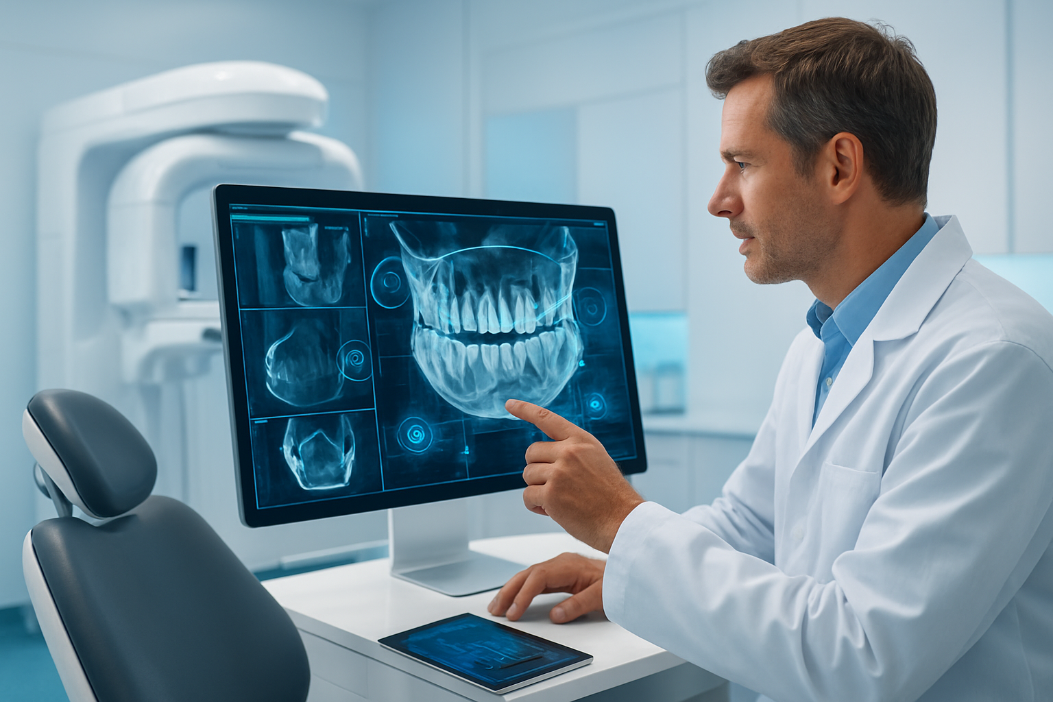
Artificial intelligence has fundamentally changed how we identify and map anatomical structures in CBCT imaging. Traditional manual identification required dentists and radiologists to spend countless hours marking anatomical landmarks, outlining nerve pathways, and identifying critical structures like the mandibular canal or maxillary sinuses. This process was not only time-consuming but also prone to human error and inconsistency between practitioners.
Modern AI systems can now automatically segment and identify dozens of anatomical structures within seconds of scanning completion. These systems use deep learning algorithms trained on thousands of annotated CBCT scans to recognize patterns and variations in bone density, tissue contrast, and spatial relationships. The technology has reached a level of accuracy that often surpasses human performance in many standardized tasks.
The most sophisticated AI platforms can identify over 40 distinct anatomical structures in a single CBCT scan. These include major landmarks like the inferior alveolar nerve canal, mental foramen, incisive canal, maxillary sinuses, nasal cavity, and temporomandibular joint components. The software also recognizes smaller structures that might be overlooked during manual analysis, such as accessory canals, anatomical variations, and subtle bone architecture changes.
Neural networks excel at recognizing complex three-dimensional relationships between structures. They can trace the complete path of the mandibular canal through varying bone densities and around pathological changes. This capability proves invaluable when planning implant placement near critical structures or when assessing the relationship between impacted teeth and adjacent anatomy.
Machine learning algorithms continue improving through exposure to diverse patient populations and anatomical variations. Systems trained on datasets from multiple geographic regions and ethnic backgrounds demonstrate better accuracy across different patient demographics. This diversity in training data helps the AI recognize uncommon anatomical variants that might confuse less sophisticated systems.
The speed of automated identification creates new workflow possibilities. Practitioners can review automatically generated anatomical maps immediately after scanning, allowing for real-time treatment planning discussions with patients. This immediate feedback transforms the patient experience and enables more efficient scheduling of follow-up procedures.
Color-coded anatomical maps generated by AI systems provide clear visual references that enhance communication between specialists. When referring patients to oral surgeons or orthodontists, practitioners can share detailed anatomical annotations that ensure consistent understanding of critical structures and their relationships. This standardized communication reduces the risk of misinterpretation and improves treatment coordination.
Quality assurance features built into modern AI systems flag unusual anatomical findings or areas where automatic identification may be uncertain. These alerts prompt manual review of specific regions, combining the efficiency of automation with the critical thinking skills of experienced practitioners. The hybrid approach maximizes both speed and accuracy in clinical practice.
Integration with existing practice management systems allows automated anatomical data to flow seamlessly into treatment planning workflows. Patient records automatically populate with detailed anatomical information, creating comprehensive documentation that supports clinical decision-making and legal compliance requirements.
Advanced systems can adapt their identification parameters based on patient age, gender, and clinical history. Pediatric patients present different anatomical proportions and developmental stages that require specialized recognition algorithms. Similarly, elderly patients may have anatomical changes related to bone remodeling or tooth loss that require different identification criteria.
Research continues expanding the range of identifiable structures. Emerging algorithms can recognize soft tissue boundaries, salivary glands, lymph nodes, and vascular structures that weren’t visible or distinguishable in earlier AI systems. This expanded recognition capability opens new diagnostic possibilities and treatment planning applications.
The technology also excels at identifying anatomical structures in challenging cases where traditional manual identification might be difficult or uncertain. Patients with previous trauma, surgical alterations, or congenital anomalies present complex anatomical relationships that AI systems can often navigate more consistently than human operators working under time pressure.
Pathology Detection and Classification Systems
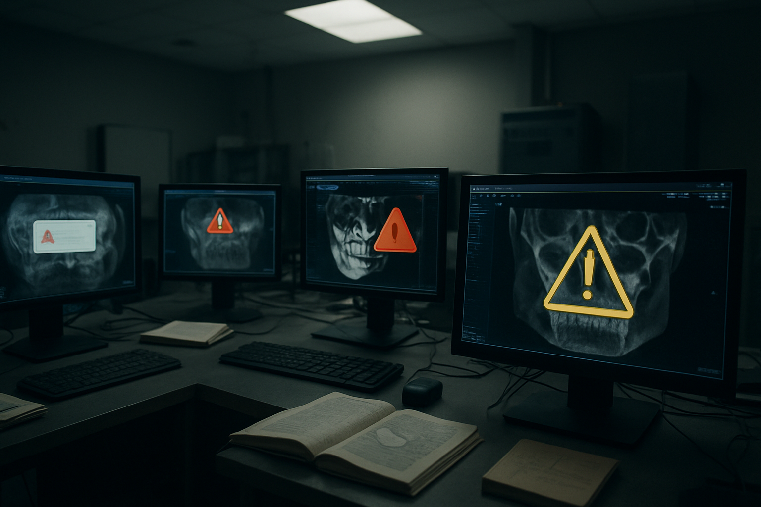
AI-powered pathology detection represents perhaps the most clinically significant advancement in CBCT interpretation. These systems can identify subtle changes in bone density, unusual growth patterns, and early signs of pathological processes that might escape detection during routine visual examination. The technology acts as a highly sensitive screening tool that ensures pathological findings receive appropriate attention and follow-up care.
Cystic lesions represent one area where AI detection has shown remarkable accuracy. Traditional identification of cysts and other radiolucent lesions often depends on the examiner’s experience and attention to detail. AI systems can detect small cystic changes measuring just a few millimeters in diameter, often before they become clinically apparent. The software analyzes bone density patterns, lesion margins, and surrounding tissue changes to classify different types of cystic pathology.
Modern classification algorithms can distinguish between various types of odontogenic cysts, including dentigerous cysts, radicular cysts, and keratocystic odontogenic tumors. Each pathology type presents distinct radiographic characteristics that AI systems learn to recognize through extensive training on confirmed cases. This classification capability helps practitioners prioritize cases requiring immediate attention versus those suitable for monitoring over time.
Periodontal bone loss detection has been revolutionized by AI analysis of subtle density changes around tooth roots. Traditional radiographic assessment of periodontal disease often underestimates the extent of bone loss, particularly in early stages. CBCT imaging combined with AI analysis can detect minimal bone density changes that precede obvious radiographic signs of periodontal destruction.
The technology quantifies bone loss with precision that exceeds human visual assessment capabilities. AI systems can measure bone height changes as small as 0.1 millimeters and track progression over time through comparative analysis of sequential scans. This level of precision enables earlier intervention and more accurate monitoring of periodontal therapy outcomes.
Impacted tooth complications receive special attention from AI pathology detection systems. Software can identify signs of root resorption on adjacent teeth, follicular enlargement around impacted crowns, and early signs of cystic transformation. These findings often remain subtle on two-dimensional radiographs but become clearly detectable when AI analyzes three-dimensional CBCT data.
Temporomandibular joint pathology detection represents another area of significant AI advancement. Joint space narrowing, condylar remodeling, erosive changes, and osteophyte formation can all be automatically detected and quantified. The software compares bilateral joint anatomy to identify asymmetries that might indicate pathological changes requiring treatment.
Root fracture identification has benefited enormously from AI analysis capabilities. Vertical root fractures often present subtle radiographic signs that can be missed during routine examination. AI systems can detect minute changes in root morphology, unusual radiolucent lines, and density variations that suggest fracture presence. This early detection capability can prevent unnecessary endodontic treatment and guide appropriate extraction decisions.
Malignancy screening represents perhaps the most critical application of AI pathology detection. While primary oral malignancies remain relatively rare, the consequences of missed diagnosis are severe. AI systems trained on large datasets of confirmed malignant lesions can identify suspicious changes in bone architecture, unusual growth patterns, and asymmetric remodeling that might indicate neoplastic processes.
The software flags cases requiring immediate specialist referral based on specific radiographic criteria associated with malignant transformation. Features like cortical destruction, irregular borders, rapid growth patterns, and associated soft tissue changes all contribute to risk assessment algorithms. This automated screening ensures that suspicious cases receive prompt attention even when practitioners may not initially recognize concerning features.
False positive rates in AI pathology detection continue improving as training datasets expand and algorithms become more sophisticated. Modern systems achieve sensitivity levels above 90% for most common pathologies while maintaining specificity rates that minimize unnecessary patient anxiety and redundant testing. The balance between detecting pathology and avoiding false alarms requires careful calibration based on clinical outcomes data.
Integration of AI pathology detection with electronic health records creates comprehensive screening protocols that track patient risk factors and recommend appropriate follow-up intervals. Patients with previous pathology, family history of oral cancer, or other risk factors receive enhanced screening attention from AI systems programmed to recognize patterns associated with increased disease probability.
Multi-institutional validation studies continue demonstrating the reliability of AI pathology detection across different scanner types, imaging protocols, and patient populations. This evidence base supports clinical adoption and provides practitioners with confidence in AI-generated findings. Regulatory approval processes are incorporating these validation studies to establish standards for AI-assisted diagnosis in dental practice.
Treatment Planning Optimization Tools
AI-driven treatment planning optimization represents the practical culmination of automated structure identification and pathology detection capabilities. These tools synthesize anatomical information, pathology findings, and treatment objectives to generate comprehensive plans that optimize clinical outcomes while minimizing patient risk and treatment time.
Implant planning has been completely transformed by AI optimization algorithms. Traditional implant planning required practitioners to manually assess bone volume, identify critical structures, and determine optimal implant positions through trial and error placement of virtual implants. Modern AI systems can evaluate thousands of potential implant positions within seconds, considering bone density distributions, anatomical constraints, and prosthetic requirements simultaneously.
The software automatically identifies the largest diameter and longest length implants that can be safely placed while maintaining appropriate distances from critical structures. Safety margins around the mandibular canal, mental foramen, maxillary sinuses, and adjacent tooth roots are automatically maintained according to established clinical guidelines or customizable practice preferences.
Bone density analysis plays a crucial role in AI-driven implant planning. The software maps bone density throughout the planned implant site and recommends drilling protocols, implant surface characteristics, and healing periods based on local bone quality. Areas of low bone density receive special attention with recommendations for site development procedures or alternative treatment approaches.
Prosthetically-driven implant planning represents a significant advancement enabled by AI optimization. The software considers the final restoration design and works backward to determine ideal implant positions that will support optimal prosthetic outcomes. Crown contours, emergence profiles, and occlusal requirements all influence the AI recommendations for implant placement.
Multi-implant cases benefit enormously from AI optimization capabilities. The software can simultaneously position multiple implants while maintaining appropriate inter-implant distances, parallel orientations, and optimal distribution of occlusal forces. Complex full-arch cases that previously required extensive manual planning can now be optimized automatically with consideration for immediate loading protocols and temporary restoration requirements.
Orthodontic treatment planning has embraced AI optimization for complex case management. Software can predict tooth movement patterns, identify potential complications, and recommend treatment sequences that minimize treatment time and patient discomfort. The three-dimensional analysis of root positions, bone architecture, and airway dimensions provides comprehensive information that supports optimal orthodontic outcomes.
Impacted tooth extraction planning benefits significantly from AI-assisted approach optimization. The software can model different surgical access routes, predict complications based on root morphology and adjacent anatomy, and recommend techniques that minimize surgical trauma. Proximity to critical structures like the inferior alveolar nerve receives special attention with recommendations for nerve repositioning or alternative approaches when indicated.
Endodontic treatment planning has been enhanced by AI analysis of root canal anatomy and periapical pathology. Complex root morphologies with unusual canal configurations can be mapped automatically, and treatment difficulty can be assessed objectively. Cases likely to require specialist referral can be identified early, improving patient outcomes and practice efficiency.
Periodontal regeneration procedures benefit from AI-assisted treatment planning that considers bone defect morphology, root surface area, and anatomical factors affecting regenerative potential. The software can predict likely outcomes for different regenerative approaches and recommend techniques most likely to achieve optimal results based on defect characteristics.
Surgical guide design represents a practical application where AI optimization directly impacts clinical procedures. Automatically generated surgical guides incorporate safety margins, optimal access angles, and depth controls that ensure accurate transfer of virtual planning to the surgical site. The precision of AI-generated guides often exceeds manually designed alternatives while requiring significantly less design time.
Risk assessment algorithms integrated into AI planning tools evaluate patient-specific factors that might influence treatment outcomes. Medical history, medications, smoking status, and previous treatment responses all contribute to risk calculations that influence treatment recommendations. High-risk patients receive modified protocols designed to optimize success rates while minimizing complications.
Treatment sequencing optimization considers the interdependence of different procedures and recommends timing that maximizes overall treatment efficiency. Complex cases involving orthodontics, implant placement, and prosthetic restoration can be sequenced optimally to minimize total treatment time while ensuring optimal outcomes at each stage.
Cost optimization features help practices balance clinical excellence with economic considerations. AI systems can evaluate different treatment alternatives and provide cost-benefit analyses that consider success rates, longevity expectations, and patient satisfaction outcomes. This information supports informed consent discussions and helps patients make educated treatment decisions.
Patient communication tools integrated with AI treatment planning generate visual presentations that clearly explain recommended procedures and expected outcomes. Three-dimensional animations, before-and-after predictions, and alternative treatment comparisons help patients understand complex treatment plans and make informed decisions about their care.
Quality assurance protocols built into AI planning systems flag potential issues or unusual recommendations that require human review. Cases outside normal parameters receive special attention to ensure that AI recommendations align with clinical judgment and best practices. This safety net maintains the balance between automation efficiency and clinical oversight.
Continuous learning algorithms improve treatment planning recommendations based on documented outcomes from completed cases. Practices using AI planning systems contribute anonymized outcome data that helps refine algorithms and improve future recommendations. This feedback loop ensures that AI tools continue evolving based on real-world clinical results.
Integration with CAD/CAM systems creates seamless workflows from diagnosis through final restoration delivery. Treatment plans generated by AI systems can directly drive manufacturing processes for surgical guides, temporary restorations, and final prosthetic components. This integration eliminates manual data transfer steps and reduces the potential for errors in complex treatment sequences.
Research applications of AI treatment planning tools are advancing understanding of optimal treatment protocols across different patient populations. Large-scale analysis of treatment outcomes correlated with AI planning parameters provides evidence for best practices and helps identify factors that most significantly influence treatment success. This research contributes to the ongoing refinement of clinical protocols and treatment guidelines.
Current Limitations and Technical Challenges
Data Quality Requirements for Optimal Performance
The effectiveness of artificial intelligence systems in CBCT interpretation hinges critically on the quality and characteristics of the data used to train these algorithms. Understanding these requirements reveals why many AI implementations fall short of their promised potential in real-world dental practice settings.
High-quality CBCT data for AI training demands exceptional image resolution and consistent scanning protocols. The algorithms require images with sufficient voxel resolution to distinguish between critical anatomical structures. Most successful AI models work best with voxel sizes ranging from 0.1 to 0.4 millimeters, though specific applications may require even finer resolution. When practices use lower resolution settings to reduce radiation exposure or scanning time, the resulting images may lack the detail necessary for accurate AI analysis.
Standardization across different CBCT machines presents another major challenge. Each manufacturer’s equipment produces images with slightly different characteristics in terms of contrast, brightness, noise patterns, and artifact presentation. An AI system trained primarily on data from one manufacturer’s machines may struggle when analyzing images from different equipment. This manufacturer-specific bias can lead to inconsistent performance across practices using various CBCT systems.
The volume of training data required for robust AI performance is staggering. Effective deep learning models typically need thousands or tens of thousands of annotated cases to achieve clinical-grade accuracy. Each image must be carefully labeled by experienced radiologists or oral and maxillofacial surgeons, a process that requires significant time and expertise. The annotation process becomes even more complex when dealing with pathological cases, where expert consensus may be required to establish ground truth labels.
Patient diversity in training datasets directly impacts AI performance across different populations. Most AI systems show reduced accuracy when analyzing images from patients whose demographic characteristics differ significantly from the training population. Age-related changes in bone density and anatomy can confuse AI algorithms trained primarily on younger patients. Similarly, systems trained predominantly on one ethnic group may struggle with anatomical variations common in other populations.
Image quality variations due to patient factors create additional challenges. Patient movement during scanning, metal artifacts from dental restorations, and beam hardening artifacts can significantly degrade image quality. AI systems must be trained to handle these real-world imperfections, yet many research datasets consist primarily of high-quality images that may not represent typical clinical conditions.
The temporal aspect of data collection also affects AI performance. Dental conditions evolve over time, and AI systems need training data that captures this progression. However, longitudinal datasets with multiple timepoints for the same patients are relatively rare and expensive to collect. This limitation affects the ability of AI systems to track disease progression or treatment outcomes over time.
Data preprocessing requirements add another layer of complexity. Raw CBCT data often requires extensive preprocessing before AI analysis, including noise reduction, artifact correction, and intensity normalization. Different preprocessing approaches can significantly impact AI performance, yet there’s limited consensus on optimal preprocessing protocols. Practices must ensure their preprocessing pipeline matches the requirements of their chosen AI system, which may necessitate additional software and training.
The challenge of rare pathology representation in training datasets cannot be understated. While common conditions like caries and periodontal disease are well-represented in most datasets, rare tumors, developmental anomalies, and unusual presentations may have insufficient examples for effective AI training. This leads to systems that perform well on common cases but may miss or misclassify rare but clinically significant findings.
Quality control measures for training data require ongoing attention. Even expertly annotated datasets may contain errors or inconsistencies that can propagate through AI training. Regular auditing of training data quality and inter-rater reliability assessments are essential but resource-intensive processes that many AI development efforts skimp on.
The geographic and institutional bias in training datasets poses another significant challenge. Most publicly available CBCT datasets come from academic medical centers in developed countries. These images may not represent the full spectrum of dental conditions and anatomical variations seen in different geographic regions or healthcare settings. AI systems trained on such datasets may show reduced performance when deployed in different clinical environments.
Data security and privacy requirements complicate the collection and sharing of high-quality training datasets. HIPAA compliance and international privacy regulations limit the ability to aggregate large, diverse datasets from multiple institutions. De-identification of CBCT images while preserving clinically relevant information requires sophisticated techniques and adds to data preparation costs.
The dynamic nature of CBCT technology itself creates moving targets for AI development. As scanning protocols improve and new reconstruction algorithms emerge, AI systems may need retraining on updated datasets. This creates an ongoing requirement for fresh training data and model updates that many practices are unprepared to handle.
Validation dataset quality requirements are equally stringent. AI systems need separate, high-quality datasets for performance validation that truly represent the intended use population. Many AI systems show impressive performance on their training datasets but fail to maintain this performance when tested on independent validation data from different institutions or patient populations.
Integration Barriers with Existing Practice Management Systems
The seamless integration of AI-powered CBCT interpretation tools with existing practice management systems represents one of the most significant practical challenges facing dental practices today. The complexity of modern dental practice software ecosystems creates multiple points of potential friction that can undermine the effectiveness of AI implementations.
Practice management systems in dentistry have evolved into comprehensive platforms that handle scheduling, billing, patient records, treatment planning, and imaging. These systems often use proprietary data formats and communication protocols that were designed before AI integration became a consideration. Adding AI capabilities to this existing infrastructure requires careful planning and often expensive custom development work.
The diversity of practice management software creates a fragmentation problem for AI developers. Major systems like Dentrix, Eaglesoft, Open Dental, and Practice Works each have their own APIs, data structures, and integration requirements. AI companies must either develop multiple integrations for different systems or limit their market reach to practices using specific software platforms. This fragmentation increases development costs and limits the adoption of AI solutions.
Data format compatibility issues plague many integration attempts. CBCT images are typically stored in DICOM format, but different practice management systems may handle DICOM data differently. Some systems store images in proprietary formats or compressed versions that may not be suitable for AI analysis. Converting between formats can introduce quality loss or metadata corruption that affects AI performance.
Network infrastructure limitations in many dental practices create additional integration challenges. AI processing often requires significant bandwidth for uploading large CBCT datasets to cloud-based processing services. Many practices operate with basic internet connections that cannot efficiently handle the data transfer requirements of modern AI systems. Local processing solutions may require hardware upgrades that practices are reluctant to invest in.
User authentication and access control systems present complex integration challenges. Practice management systems typically have their own user authentication mechanisms, and integrating AI tools while maintaining security and compliance requires careful coordination. Single sign-on solutions may not be available or may require expensive customization to accommodate AI system requirements.
Workflow integration represents perhaps the most challenging aspect of AI implementation. Dental practices have established workflows for image acquisition, interpretation, and treatment planning that have been refined over years of practice. Introducing AI tools that require additional steps or changes to existing workflows often meets resistance from staff who are comfortable with current procedures.
The timing and sequence of AI analysis within practice workflows require careful consideration. Some AI systems require images to be processed immediately after acquisition, while others can analyze images retrospectively. Practices must determine when in their workflow AI analysis should occur and how the results should be presented to clinicians. Poor timing can disrupt patient flow or create bottlenecks in busy practices.
Training requirements for staff represent a significant barrier to successful integration. Practice management systems are already complex, and adding AI capabilities increases the learning curve for dental professionals and staff. Many practices lack the time and resources for comprehensive training programs, leading to underutilization of AI capabilities or user frustration.
Data synchronization between AI systems and practice management platforms creates ongoing technical challenges. Patient demographics, treatment histories, and imaging metadata must be kept synchronized across systems to ensure accurate AI analysis and proper record keeping. Any discrepancies in patient data can lead to mismatched analyses or compliance issues.
Backup and disaster recovery procedures become more complex with AI integration. Practices must ensure that AI-generated analyses and recommendations are properly backed up along with other patient data. Recovery procedures must account for AI system dependencies and may require coordination with external AI service providers.
The cost structure of integrated AI solutions often doesn’t align with practice budgets and billing systems. Many AI systems use subscription-based pricing models that may not match the fee-for-service structure of dental practice revenue. Practices struggle to determine the return on investment for AI systems, especially when integration costs are factored in.
Version control and update management become critical issues with integrated AI systems. Practice management systems typically have their own update schedules and may not be compatible with frequent AI model updates. Ensuring that all system components remain compatible while allowing for AI improvements requires careful coordination.
Legacy system support presents ongoing challenges for AI integration. Many dental practices use older practice management systems that lack modern integration capabilities. These systems may require expensive upgrades or complete replacements to accommodate AI integration, costs that many practices cannot justify.
Vendor relationship management becomes more complex with AI integration. Practices must now coordinate with multiple vendors including their practice management system provider, CBCT equipment manufacturer, and AI solution provider. When technical issues arise, determining responsibility and coordinating solutions across multiple vendors can be frustrating and time-consuming.
Performance optimization for integrated systems requires ongoing attention. AI processing can be computationally intensive and may slow down other practice management functions if not properly managed. Practices must monitor system performance and may need to upgrade hardware or adjust workflows to maintain optimal operation.
Data portability concerns arise when practices want to change AI providers or practice management systems. Patient data and AI-generated analyses must be exportable in standard formats to avoid vendor lock-in situations. Many current integration solutions lack adequate data portability features, creating long-term risks for practices.
Customization requirements for practice-specific workflows often exceed the capabilities of standard integration solutions. Each practice may have unique requirements based on their patient population, specialties, and operational preferences. AI integration solutions that cannot accommodate these variations may force practices to change their workflows rather than enhancing them.
Regulatory Compliance and Approval Processes
The regulatory landscape surrounding AI applications in medical imaging presents a complex web of requirements that significantly impact the development and deployment of CBCT interpretation systems. Understanding these regulatory frameworks is essential for both AI developers and dental practices considering implementation.
The FDA’s approach to regulating AI-based medical devices has evolved significantly in recent years. Traditional medical device regulations were designed for static devices with fixed functionality, but AI systems can learn and change over time, creating new regulatory challenges. The FDA has developed a framework for Software as Medical Device (SaMD) that applies to AI systems used for diagnostic purposes, including CBCT interpretation tools.
Classification of AI systems under FDA regulations depends on their intended use and risk level. AI tools that provide diagnostic recommendations or automated detection of pathology are typically classified as Class II medical devices, requiring 510(k) clearance before marketing. This process involves demonstrating substantial equivalence to existing approved devices and providing clinical validation data. The timeline for 510(k) clearance can range from several months to over a year, creating significant barriers for AI companies seeking to enter the market.
Clinical validation requirements for AI systems are particularly stringent. The FDA requires robust clinical studies demonstrating that AI systems perform as well as or better than existing standard of care. These studies must include diverse patient populations and multiple clinical sites to demonstrate generalizability. The cost and time required for these studies often exceed the resources available to smaller AI companies, leading to market concentration among larger organizations.
The challenge of demonstrating AI system performance in regulatory submissions goes beyond simple accuracy metrics. The FDA requires comprehensive analyses of system failures, bias assessments, and clear documentation of system limitations. AI companies must provide detailed information about training datasets, validation methodologies, and ongoing monitoring procedures. This documentation burden requires significant regulatory expertise that many AI startups lack.
Post-market surveillance requirements for AI systems add ongoing compliance obligations. The FDA expects manufacturers to monitor AI system performance in real-world use and report adverse events or performance degradation. For AI systems that continue to learn from new data, manufacturers must have procedures for monitoring and controlling algorithm changes. These requirements create ongoing costs and operational burdens that must be factored into business models.
International regulatory harmonization efforts are still developing for AI medical devices. While the FDA leads in many areas, regulatory agencies in Europe, Canada, and other countries have their own requirements for AI system approval. The EU’s Medical Device Regulation (MDR) includes specific provisions for AI systems, but the interpretation and implementation of these requirements continue to evolve. This regulatory fragmentation means AI companies seeking global markets must navigate multiple approval processes with different requirements.
Quality management system requirements for AI medical devices extend beyond traditional software development practices. ISO 13485 certification is typically required for medical device manufacturers, but this standard was developed before AI became prevalent. Companies must adapt quality management practices to address AI-specific risks such as training data quality, model validation, and algorithm bias. This adaptation requires significant investment in quality systems and personnel training.
Data governance requirements for AI systems create additional compliance challenges. Training and validation datasets must meet strict quality and documentation standards. Patient consent for use of imaging data in AI development may be required, and de-identification procedures must comply with HIPAA and other privacy regulations. The international transfer of training data may be restricted by data localization requirements in some jurisdictions.
Clinical evidence requirements for AI systems differ significantly from traditional diagnostic devices. Randomized controlled trials may not be appropriate for AI validation, leading to reliance on retrospective studies and real-world evidence. However, regulatory agencies are still developing standards for what constitutes adequate clinical evidence for AI systems. This uncertainty makes it difficult for companies to plan clinical validation strategies and budget appropriately.
Labeling and user training requirements for AI medical devices must address the unique characteristics of AI systems. Users must understand system limitations, appropriate use cases, and failure modes. The FDA requires clear labeling about AI system performance characteristics and recommends user training programs. These requirements add to the cost and complexity of AI system deployment in clinical practice.
Risk management procedures for AI systems must address novel failure modes not seen in traditional medical devices. Algorithm bias, adversarial attacks, and data drift represent new categories of risks that must be identified, assessed, and mitigated. ISO 14971 risk management principles apply to AI devices, but companies must adapt these principles to address AI-specific risks. This adaptation requires deep understanding of both AI technology and risk management practices.
Cybersecurity requirements for AI medical devices have become increasingly stringent. The FDA’s cybersecurity guidance requires manufacturers to address security throughout the device lifecycle, from design through deployment and maintenance. AI systems may be particularly vulnerable to certain types of cyberattacks, such as adversarial examples that can fool image recognition algorithms. Manufacturers must implement robust cybersecurity measures and maintain them throughout the product lifecycle.
Software lifecycle processes for AI systems must accommodate the iterative nature of AI development. Traditional waterfall development models may not be suitable for AI systems that require frequent retraining and model updates. Agile development practices must be adapted to meet regulatory requirements for documentation, validation, and change control. This adaptation requires careful planning and may slow down development cycles compared to non-regulated software.
Interoperability requirements for AI medical devices add another layer of regulatory complexity. The FDA encourages the development of interoperable medical devices that can work with multiple platforms and systems. For AI CBCT interpretation tools, this means ensuring compatibility with different CBCT manufacturers, practice management systems, and imaging standards. Meeting interoperability requirements while maintaining device safety and effectiveness requires careful design and extensive testing.
Intellectual property considerations intersect with regulatory requirements in complex ways. AI algorithms may be protected by patents or trade secrets, but regulatory submissions require detailed disclosure of system functionality. Companies must balance intellectual property protection with regulatory transparency requirements. Patent freedom to operate analyses become more complex for AI systems that may incorporate multiple patented technologies.
Reimbursement pathway development represents a parallel regulatory challenge. While FDA approval may be necessary for marketing AI devices, separate approval from the Centers for Medicare & Medicaid Services (CMS) may be required for reimbursement. The lack of specific billing codes for AI-assisted diagnosis creates barriers to practice adoption even after regulatory approval. Developing reimbursement pathways requires engagement with multiple stakeholders and may take years to complete.
State-level regulatory requirements add another layer of complexity for AI medical devices. While the FDA regulates medical devices at the federal level, states may have additional requirements for software used in clinical practice. Some states require software registration or impose specific liability requirements that affect AI system deployment. Companies must understand and comply with state-level requirements in each market they serve.
Professional liability considerations affect both AI developers and end users. Malpractice insurance for healthcare providers may not clearly cover AI-assisted diagnosis, creating potential gaps in coverage. AI companies must consider product liability exposure and may need specialized insurance products. Clear agreements about liability allocation between AI providers and healthcare facilities are essential but often difficult to negotiate.
Continuing education requirements for healthcare providers using AI systems vary by state and professional organization. Some jurisdictions require specific training in AI technology before clinicians can use AI diagnostic tools. These requirements affect the market adoption of AI systems and may influence product design decisions. AI companies may need to develop and maintain training programs to support regulatory compliance by their customers.
The rapid pace of AI technology development creates challenges for regulatory frameworks designed for more stable technologies. Regulatory guidance documents may quickly become outdated as AI technology advances. Companies must stay current with evolving regulatory interpretations and may need to adapt their products to meet changing requirements. This regulatory uncertainty increases development costs and risks for AI companies.
Pre-submission meetings with regulatory agencies can help clarify requirements but require significant preparation and expertise. The FDA offers Q-Sub meetings for AI device developers to discuss regulatory pathways before formal submissions. These meetings can provide valuable guidance but require comprehensive documentation packages and may involve long wait times for scheduling. Smaller companies may lack the resources to effectively utilize these pre-submission opportunities.
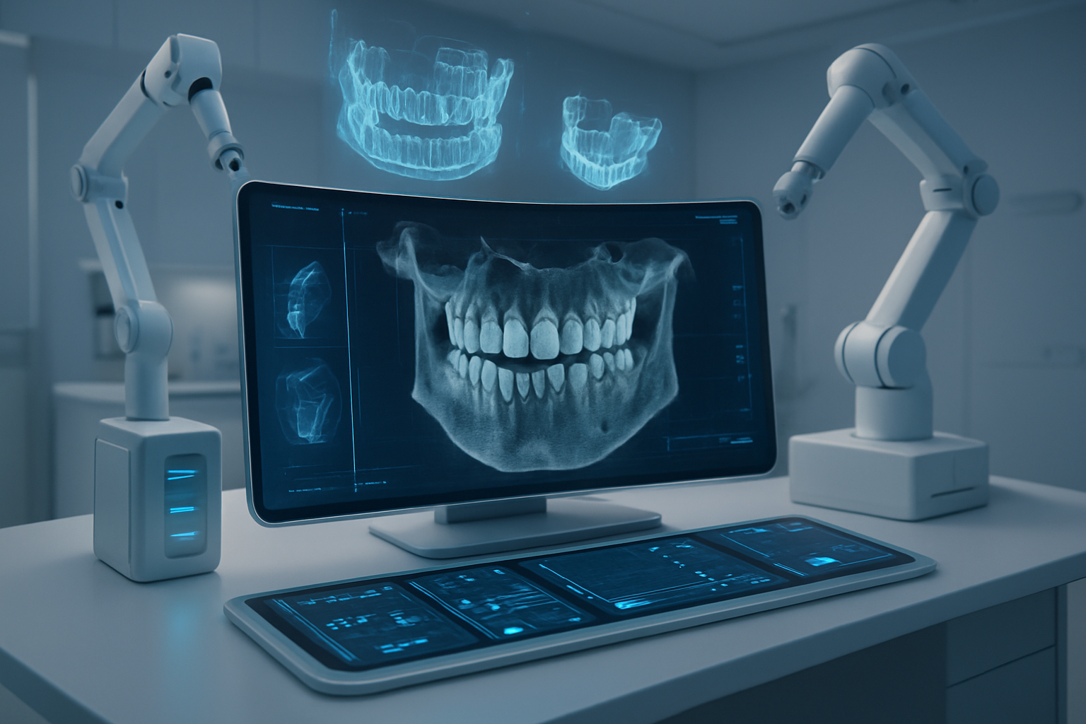
Emerging Technologies Shaping Tomorrow’s CBCT Interpretation
Advanced Neural Networks for Complex Pattern Recognition
The evolution of neural network architectures represents one of the most significant advances in CBCT interpretation technology. These sophisticated systems go far beyond traditional machine learning approaches, offering unprecedented capabilities for recognizing complex anatomical patterns and pathological conditions that might escape even experienced clinicians.
Convolutional Neural Networks (CNNs) have become the backbone of modern CBCT analysis, but their current incarnations represent just the beginning. Next-generation deep learning models incorporate residual connections, attention mechanisms, and transformer architectures that dramatically improve pattern recognition accuracy. These networks can simultaneously analyze multiple anatomical regions while maintaining awareness of spatial relationships between structures.
One particularly promising development involves multi-scale feature extraction networks that analyze CBCT data at various resolution levels simultaneously. These systems excel at detecting both macro-level anatomical variations and micro-level pathological changes within the same scan. The architecture essentially creates a hierarchy of analysis, where high-level networks identify major anatomical landmarks while specialized sub-networks focus on specific regions of interest.
Graph neural networks represent another breakthrough in CBCT interpretation. These networks model anatomical structures as interconnected nodes, allowing the AI to understand relationships between different dental and maxillofacial components. This approach proves especially valuable for complex cases involving multiple missing teeth, extensive bone loss, or anatomical anomalies where traditional spatial analysis might fall short.
The integration of generative adversarial networks (GANs) into CBCT interpretation opens new possibilities for data augmentation and synthetic image generation. These networks can create realistic CBCT images representing various pathological conditions, helping train more robust diagnostic models. More importantly, GANs can enhance low-quality scans by predicting and filling in missing information, effectively improving image resolution and clarity for better diagnostic accuracy.
Attention-based neural networks mark a paradigm shift in how AI systems process CBCT data. These networks dynamically focus on relevant regions during analysis, mimicking how human radiologists visually scan images. The attention mechanism allows the network to build hierarchical representations of important features while suppressing irrelevant background information. This approach significantly improves diagnostic accuracy for subtle pathological conditions that might otherwise be overlooked.
Self-supervised learning represents a game-changing approach for CBCT interpretation. These networks learn meaningful representations from unlabeled data by solving pretext tasks, such as predicting masked portions of images or identifying spatial relationships between image patches. This approach addresses the persistent challenge of limited labeled training data in medical imaging while creating more generalizable models.
Ensemble learning methods combine multiple neural network architectures to create more robust and accurate diagnostic systems. These approaches leverage the strengths of different network types, such as combining CNNs for spatial analysis with recurrent neural networks for temporal pattern recognition in follow-up scans. The ensemble approach significantly reduces false positive and false negative rates while improving overall diagnostic confidence.
Few-shot learning capabilities enable neural networks to adapt quickly to new pathological conditions or anatomical variations with minimal training examples. This flexibility proves crucial in dental practice, where practitioners encounter rare conditions or unique anatomical presentations that weren’t adequately represented in the original training data.
The development of explainable AI mechanisms within neural networks addresses the critical need for transparency in medical diagnosis. These systems provide visual explanations of their decision-making process, highlighting specific regions and features that contributed to diagnostic conclusions. This transparency builds trust with clinicians and enables better integration of AI recommendations into treatment planning workflows.
Continual learning architectures allow neural networks to continuously improve their performance as they encounter new cases. These systems can update their knowledge base without forgetting previously learned information, creating AI assistants that become more accurate and reliable over time within specific practice environments.
Cloud-Based AI Solutions Enabling Remote Analysis
Cloud computing infrastructure has revolutionized the accessibility and scalability of AI-powered CBCT interpretation, transforming sophisticated diagnostic capabilities from expensive on-premise solutions into accessible, subscription-based services. This shift democratizes advanced imaging analysis, making cutting-edge AI tools available to practices of all sizes regardless of their local computing resources.
The architecture of cloud-based CBCT analysis platforms leverages distributed computing power to process complex three-dimensional imaging data that would overwhelm traditional desktop workstations. Modern cloud platforms utilize GPU clusters specifically optimized for deep learning inference, enabling real-time processing of high-resolution CBCT scans. These systems can handle multiple concurrent analyses while maintaining sub-minute processing times for standard diagnostic tasks.
Edge computing integration represents a hybrid approach that combines local processing capabilities with cloud-based AI models. This architecture addresses concerns about data transmission times and internet connectivity by performing initial image preprocessing locally while leveraging cloud resources for complex diagnostic analysis. The result is faster overall processing times and improved reliability for practices in areas with limited internet bandwidth.
Federated learning platforms enable collaborative AI development without compromising patient privacy. These systems allow multiple dental practices to contribute to model training while keeping patient data on local servers. The AI model travels between participating sites, learning from each dataset without centralizing sensitive information. This approach creates more robust diagnostic models trained on diverse patient populations while maintaining HIPAA compliance and data security.
Containerized AI services provide unprecedented flexibility in deploying and scaling CBCT interpretation tools. Docker and Kubernetes technologies enable rapid deployment of specialized AI models for specific diagnostic tasks, allowing practices to access the most appropriate tools for their patient populations. These containers can be dynamically allocated based on demand, ensuring optimal resource utilization and cost efficiency.
Application Programming Interface (API) integration enables seamless connectivity between existing practice management systems and cloud-based AI services. Modern APIs support real-time data exchange, allowing CBCT scans to be automatically uploaded for analysis and results to be directly integrated into patient records. This streamlined workflow eliminates manual data transfer and reduces the potential for errors in the diagnostic process.
Microservices architecture breaks down complex AI analysis into specialized, independent services that can be combined as needed. For example, one microservice might specialize in airway analysis while another focuses on periodontal assessment. This modular approach allows practices to customize their AI toolkit based on specific needs while maintaining cost efficiency by only paying for utilized services.
Auto-scaling capabilities ensure that cloud-based AI platforms can handle varying workloads without performance degradation. During peak usage periods, additional computing resources are automatically allocated to maintain processing speeds. This elasticity eliminates the need for practices to invest in expensive hardware to handle occasional spikes in imaging volume.
Global content delivery networks (CDNs) optimize the performance of cloud-based CBCT analysis by distributing processing capabilities across multiple geographic regions. This infrastructure reduces latency and ensures reliable access to AI services regardless of the practice’s location. Advanced CDNs can intelligently route analysis requests to the nearest available processing center for optimal performance.
Blockchain technology integration provides secure, immutable records of AI analysis results and processing history. This technology ensures that diagnostic reports cannot be tampered with and provides a clear audit trail for quality assurance and legal purposes. Smart contracts can automate billing and service agreements while maintaining transparency and trust between service providers and dental practices.
Multi-tenancy architecture allows cloud providers to serve multiple dental practices while maintaining strict data isolation and security. Each practice’s data and analysis results remain completely separate while sharing underlying computing infrastructure. This approach maximizes cost efficiency while ensuring privacy and regulatory compliance.
Real-time collaboration features enable remote consultation and second opinions on complex CBCT cases. Cloud platforms can facilitate secure sharing of imaging data and AI analysis results between practitioners, specialists, and educational institutions. These collaboration tools support both synchronous and asynchronous consultation workflows, accommodating different scheduling needs and time zones.
Version control systems for AI models ensure that practices always have access to the latest diagnostic capabilities while maintaining the option to revert to previous versions if needed. These systems track model performance metrics and automatically alert practices to significant improvements or potential issues with updated algorithms.
Disaster recovery and data backup services provide peace of mind for practices relying on cloud-based AI analysis. Automated backup systems ensure that patient data and analysis results are protected against hardware failures or other catastrophic events. Geographically distributed backup locations provide additional security against regional disasters.
Augmented Reality Integration for Enhanced Visualization
Augmented reality technology transforms CBCT interpretation from a traditional two-dimensional analysis process into an immersive, three-dimensional diagnostic experience. This revolutionary approach overlays digital information onto real-world environments, creating intuitive interfaces that enhance diagnostic accuracy while reducing interpretation time and cognitive load.
Mixed reality headsets specifically designed for medical applications provide high-resolution displays capable of rendering complex anatomical structures with millimeter precision. These devices incorporate advanced tracking systems that maintain accurate registration between virtual CBCT data and real-world reference points, enabling seamless manipulation of three-dimensional imaging data through natural hand gestures and head movements.
Holographic projection systems represent the cutting edge of AR visualization for CBCT data. These platforms create volumetric displays that float in space, allowing multiple practitioners to simultaneously view and interact with the same three-dimensional anatomical reconstruction. The shared visualization experience enhances collaborative diagnosis and treatment planning while providing unprecedented spatial understanding of complex anatomical relationships.
Gesture recognition technology enables intuitive interaction with CBCT data without physical controllers or keyboards. Advanced computer vision algorithms track hand movements and finger positions, allowing practitioners to rotate, zoom, and section through three-dimensional anatomical structures using natural gestures. This hands-free interaction maintains sterile conditions while providing precise control over visualization parameters.
Real-time volume rendering algorithms optimize the display of CBCT data for AR environments. These systems dynamically adjust rendering quality and detail levels based on viewing distance and interaction requirements, ensuring smooth performance while maintaining diagnostic quality. Adaptive rendering techniques can emphasize pathological areas while reducing visual noise in healthy anatomical regions.
Spatial audio integration provides another dimension of information in AR-enhanced CBCT interpretation. Three-dimensional audio cues can alert practitioners to areas of interest, provide spoken diagnostic suggestions, or deliver contextual information about anatomical structures. This multi-sensory approach reduces cognitive load while improving diagnostic confidence and accuracy.
Collaborative AR platforms enable remote specialists to participate in CBCT interpretation sessions as if they were physically present. These systems synchronize AR environments across multiple locations, allowing experts to point out specific anatomical features, draw annotations, or guide less experienced practitioners through complex diagnostic procedures. This capability democratizes access to specialist expertise regardless of geographic constraints.
Digital annotation and markup tools allow practitioners to add persistent notes and measurements directly to three-dimensional CBCT reconstructions. These annotations remain visible across different viewing sessions and can be shared with other members of the treatment team. Advanced annotation systems support voice notes, sketched diagrams, and standardized diagnostic codes for comprehensive case documentation.
Template overlay systems superimpose ideal anatomical references onto patient-specific CBCT data, facilitating comparison and treatment planning. These templates can represent normal anatomical variations, ideal occlusal relationships, or planned implant positions. The overlay visualization helps identify deviations from normal anatomy and guides treatment planning decisions.
Predictive visualization capabilities project potential treatment outcomes directly onto CBCT data. These systems can simulate bone remodeling following tooth extraction, visualize planned orthodontic movements, or predict soft tissue changes after orthognathic surgery. Real-time visualization of treatment predictions enhances patient communication while improving treatment planning accuracy.
Integration with surgical navigation systems creates seamless workflows from diagnosis through treatment execution. AR platforms can display planned surgical approaches overlaid on CBCT data, then transfer this information to intraoperative navigation systems for precise implementation. This integration reduces the gap between diagnostic planning and surgical execution while improving treatment predictability.
Machine learning-powered region highlighting automatically identifies and emphasizes areas of diagnostic interest within AR-rendered CBCT data. These systems can highlight suspected pathological areas, flag anatomical anomalies, or draw attention to critical anatomical structures based on the specific diagnostic task. Dynamic highlighting adapts to practitioner behavior and diagnostic preferences over time.
Cross-platform compatibility ensures that AR-enhanced CBCT visualization works across different hardware platforms and operating systems. Cloud-based rendering services can deliver AR content to various devices, from high-end medical displays to tablet computers, making advanced visualization capabilities accessible in different clinical environments.
Eye tracking technology monitors practitioner gaze patterns during CBCT interpretation, providing insights into diagnostic decision-making processes. This data can identify overlooked anatomical areas, optimize user interface design, and provide feedback for continuing education. Advanced systems can proactively guide attention to important features based on successful diagnostic patterns.
Predictive Analytics for Treatment Outcome Forecasting
Predictive analytics represents a paradigm shift in dental treatment planning, moving beyond reactive diagnosis toward proactive outcome forecasting. These sophisticated systems analyze vast datasets of historical treatment cases, patient characteristics, and long-term follow-up data to predict treatment success rates, potential complications, and optimal therapeutic approaches for individual patients.
Longitudinal outcome modeling leverages machine learning algorithms to analyze treatment results across extended timeframes, sometimes spanning decades of patient data. These models identify patterns that correlate specific patient characteristics, treatment modalities, and clinical techniques with long-term success rates. The resulting predictions help practitioners select treatment approaches with the highest probability of favorable outcomes for each unique patient presentation.
Risk stratification algorithms automatically categorize patients based on their likelihood of experiencing specific complications or treatment failures. These systems consider multiple variables including medical history, anatomical factors, lifestyle characteristics, and genetic markers to assign risk scores for various treatment scenarios. High-risk patients can receive modified treatment protocols or increased monitoring to prevent adverse outcomes.
Biomechanical modeling systems predict how different treatment approaches will affect stress distribution, bone remodeling, and tissue healing processes. These models incorporate patient-specific anatomical data from CBCT scans with mechanical property databases to simulate treatment outcomes before implementation. Finite element analysis can predict implant stability, orthodontic movement patterns, and prosthetic longevity with remarkable accuracy.
Treatment pathway optimization algorithms recommend the most efficient sequence of therapeutic interventions for complex multidisciplinary cases. These systems consider treatment interdependencies, healing timelines, and patient preferences to create personalized treatment schedules that minimize overall treatment time while maximizing clinical outcomes. The optimization process accounts for resource availability and scheduling constraints within specific practice environments.
Complication prediction models identify patients at elevated risk for specific adverse events before treatment begins. These systems analyze historical patterns associated with surgical site infections, implant failures, delayed healing, or other complications to alert practitioners to potential risks. Early identification enables implementation of preventive measures that significantly reduce complication rates.
Healing timeline prediction helps practitioners and patients plan realistic treatment schedules based on individual healing characteristics. Machine learning models analyze patient factors that influence healing rates, including age, medical conditions, medications, and lifestyle factors. Accurate healing predictions improve patient satisfaction while enabling more efficient practice scheduling and resource allocation.
Maintenance requirement forecasting predicts the long-term care needs for different treatment modalities. These models estimate the frequency and type of maintenance visits required for implants, crowns, orthodontic appliances, and other dental treatments. This information helps patients make informed decisions about treatment options while enabling practices to plan appropriate follow-up care schedules.
Comparative effectiveness research powered by predictive analytics provides evidence-based recommendations for treatment selection. These systems analyze outcomes data across thousands of similar cases to identify which treatment approaches produce the best results for specific patient profiles. Real-world evidence supplements clinical trial data with insights from diverse patient populations and practice environments.
Quality assurance algorithms continuously monitor treatment outcomes to identify deviations from predicted results. These systems alert practitioners when actual outcomes differ significantly from predictions, triggering quality improvement processes and enabling rapid identification of technique or equipment issues. Continuous monitoring helps maintain consistent treatment quality while identifying opportunities for improvement.
Personalized maintenance protocols adapt follow-up care recommendations based on individual patient risk profiles and treatment responses. Predictive models analyze how different patients respond to various maintenance approaches, enabling customized care plans that optimize long-term treatment success. High-risk patients might receive more frequent monitoring while low-risk patients can follow extended maintenance schedules.
Treatment cost prediction models forecast the total financial investment required for complex treatment plans, including initial procedures, potential complications, and long-term maintenance requirements. These predictions help patients make informed financial decisions while enabling practices to provide accurate treatment estimates. Cost predictions account for the probability of various treatment scenarios and their associated expenses.
Integration with electronic health records enables seamless incorporation of predictive analytics into existing clinical workflows. These systems automatically extract relevant patient data to generate predictions without requiring additional data entry from clinical staff. Real-time integration ensures that predictions are always based on the most current patient information available.
Population health analytics identify trends and patterns across entire patient populations, enabling public health insights and community-based treatment planning. These systems can predict disease prevalence, treatment demand, and resource requirements for specific geographic regions or demographic groups. Population-level insights inform public policy decisions and healthcare resource allocation strategies.
Personalized AI Models Adapting to Individual Practice Patterns
The future of CBCT interpretation lies in AI systems that learn and adapt to the unique characteristics, preferences, and expertise patterns of individual dental practices. These personalized models represent a significant evolution from one-size-fits-all diagnostic tools toward intelligent assistants that become increasingly valuable as they accumulate experience within specific clinical environments.
Practice-specific learning algorithms continuously analyze diagnostic decisions, treatment preferences, and outcome patterns unique to each dental practice. These systems identify correlations between practitioner techniques, patient demographics, and treatment outcomes to develop customized diagnostic and treatment recommendation engines. The AI learns which approaches work best for each practitioner’s style and patient population, creating truly personalized clinical decision support.
Adaptive threshold systems automatically adjust diagnostic sensitivity based on individual practitioner preferences and expertise levels. These models learn from feedback patterns to optimize the balance between diagnostic sensitivity and specificity for each user. Experienced specialists might prefer higher sensitivity settings that flag subtle findings, while general practitioners might benefit from more conservative thresholds that focus on clear pathological conditions.
Workflow optimization engines study individual practice patterns to streamline CBCT interpretation processes. These systems learn how different practitioners prefer to analyze images, which diagnostic sequences they follow, and how they integrate AI recommendations into their decision-making process. The AI then adapts its interface and recommendation timing to match each practitioner’s natural workflow patterns.
Patient population modeling creates practice-specific diagnostic baselines that account for unique demographic characteristics, prevalent conditions, and treatment patterns within each practice’s patient base. These models recognize that urban practices might see different pathological patterns than rural practices, and pediatric specialists require different diagnostic criteria than oral surgeons. Localized baselines improve diagnostic accuracy while reducing false positive rates.
Continuous learning feedback loops enable AI models to improve their performance based on treatment outcomes and practitioner corrections. When practitioners override AI recommendations or modify suggested treatment plans, the system learns from these decisions to provide better recommendations in similar future cases. This feedback mechanism ensures that the AI becomes increasingly aligned with each practice’s clinical philosophy and standards.
Expertise augmentation systems recognize individual practitioner strengths and limitations to provide targeted diagnostic support. These models might offer more detailed explanations and guidance for complex cases that fall outside a practitioner’s primary area of expertise while providing streamlined recommendations for familiar conditions. The AI becomes a personalized continuing education tool that helps expand each practitioner’s diagnostic capabilities.
Practice management integration creates AI models that understand scheduling constraints, equipment limitations, and referral patterns unique to each practice environment. These systems can recommend treatment approaches that align with available resources and established workflows while suggesting referrals to trusted specialists when cases exceed in-house capabilities. The AI becomes intimately familiar with how each practice operates and makes recommendations accordingly.
Case complexity assessment algorithms learn to evaluate diagnostic challenges based on individual practitioner experience levels. These systems might flag cases as requiring specialist consultation for less experienced practitioners while providing detailed analysis tools for experts comfortable handling complex diagnoses. The AI adapts its support level to match each user’s diagnostic confidence and capabilities.
Historical case retrieval systems maintain searchable databases of previous cases specific to each practice, enabling rapid comparison with similar presentations. These systems learn which case characteristics are most relevant for different practitioners and automatically suggest similar historical cases during diagnostic workflows. Practice-specific case libraries become invaluable reference tools for clinical decision-making.
Quality metrics tracking monitors diagnostic accuracy and treatment outcomes for individual practitioners, providing personalized feedback for continuous improvement. These systems identify patterns in diagnostic accuracy, highlight areas for potential improvement, and track progress over time. The AI becomes a personal performance coach that helps practitioners refine their diagnostic skills through objective data analysis.
Collaborative learning networks enable AI models to share insights across different practices while maintaining individual customization. These systems can incorporate successful diagnostic patterns from other similar practices while preserving the unique characteristics that make each practice’s model valuable. Federated learning approaches maintain privacy while enabling collective improvement in diagnostic capabilities.
Subspecialty adaptation allows general AI models to develop specialized expertise based on practice focus areas. An orthodontic practice’s AI might become exceptionally skilled at airway analysis and growth prediction, while an oral surgery practice’s system might excel at surgical risk assessment and complications prediction. The AI develops subspecialty knowledge that matches each practice’s clinical focus.
Training and onboarding systems help new practitioners or staff members quickly adapt to practice-specific AI tools. These systems provide personalized training recommendations based on individual learning patterns and clinical experience levels. The AI becomes a mentor that helps new team members integrate seamlessly into established diagnostic workflows while maintaining practice standards.
Regulatory compliance monitoring ensures that practice-specific AI adaptations maintain appropriate diagnostic standards and regulatory requirements. These systems track AI recommendations against established guidelines while allowing for practice-specific customization within acceptable
Strategic Implementation Roadmap for Dental Practices
Assessment of Current Technology Infrastructure
Understanding where your dental practice stands technologically is the first step toward successful AI-powered CBCT implementation. Most dental practices today operate with a mix of legacy systems and newer digital solutions, creating a complex technological landscape that requires careful evaluation.
Your existing CBCT hardware forms the foundation of any AI integration strategy. Modern AI algorithms demand specific image quality standards and data formats that older equipment may not support. If your practice operates with CBCT machines manufactured before 2015, you’ll likely face compatibility challenges with advanced AI software. These older systems often produce images in proprietary formats that require conversion processes, potentially introducing artifacts that reduce AI diagnostic accuracy.
Image resolution and reconstruction algorithms play a critical role in AI performance. Contemporary AI systems perform optimally with voxel sizes of 0.3mm or smaller, field-of-view specifications that capture complete anatomical regions of interest, and reconstruction protocols that minimize beam hardening artifacts. Your current CBCT system should support multiple scanning protocols to accommodate different AI applications, from endodontic planning requiring high-resolution limited field-of-view scans to orthognathic surgery planning demanding full skull imaging.
The computing infrastructure supporting your CBCT operations needs significant evaluation. AI processing requires substantial computational power, with GPU-accelerated workstations becoming increasingly necessary for real-time analysis. Your current workstation specifications directly impact AI processing speed and accuracy. Systems with less than 16GB RAM and without dedicated graphics processing units will struggle with modern AI algorithms, creating bottlenecks that reduce practice efficiency.
Network infrastructure often becomes the overlooked limiting factor in AI implementation. CBCT files range from 50MB to several gigabytes depending on scan parameters, and AI processing may require cloud-based analysis or real-time data synchronization between workstations. Your practice’s internet bandwidth, local area network configuration, and data storage systems must accommodate these increased data flows without compromising patient care workflows.
Data storage architecture requires comprehensive review before AI implementation. Traditional practice management systems designed for 2D radiographs may lack the capacity and organization structure needed for CBCT data enhanced with AI annotations. Your current storage solution should support DICOM standards, maintain patient privacy compliance, and provide rapid access to historical scans for AI comparison algorithms.
Evaluating your practice’s DICOM infrastructure reveals critical compatibility requirements. AI systems integrate through DICOM communication protocols, requiring your existing systems to support advanced DICOM services beyond basic image transfer. DICOM Structured Reporting (SR) capabilities become essential for AI-generated findings documentation, while DICOM Worklist Management ensures seamless integration with your practice’s scheduling and patient management workflows.
Your current software ecosystem requires detailed mapping to identify integration points and potential conflicts. Practice management software, imaging software, treatment planning applications, and patient communication systems all interact with CBCT data. AI implementation success depends on these systems working harmoniously together, sharing data efficiently while maintaining individual functionality.
Security infrastructure assessment becomes paramount when introducing AI systems that may process patient data through cloud services or third-party algorithms. Your current cybersecurity measures, including firewalls, antivirus software, user access controls, and data encryption protocols, must align with healthcare data protection requirements while accommodating AI system communication needs.
Staff technical competency evaluation provides insight into training requirements and implementation timeline considerations. Your team’s comfort level with current technology directly influences how quickly they’ll adapt to AI-enhanced workflows. Practices with tech-savvy staff members can often implement AI systems more rapidly, while those requiring extensive digital literacy training may need extended implementation periods.
Budget allocation for infrastructure upgrades requires realistic assessment of current system limitations versus AI requirements. Hardware upgrades, software licensing, network improvements, and security enhancements all contribute to implementation costs. Understanding these requirements early in the planning process prevents mid-implementation surprises that could derail your AI adoption timeline.
Your practice’s patient volume and scan frequency influence infrastructure requirements significantly. High-volume practices processing dozens of CBCT scans daily require more robust computing power and storage solutions than smaller practices with occasional scanning needs. AI systems that provide real-time analysis during patient appointments demand different infrastructure specifications than batch processing solutions used for case review.
Vendor relationship evaluation helps identify potential integration pathways and support resources. Your current CBCT manufacturer may offer AI solutions specifically designed for their equipment, potentially simplifying integration but limiting algorithm choice. Independent AI vendors might provide more advanced capabilities but require more complex integration processes.
Regulatory compliance status of your current systems affects AI implementation strategies. HIPAA compliance, FDA device registrations, and state regulatory requirements must be maintained throughout AI integration. Your existing compliance framework should accommodate new AI systems without creating regulatory gaps that could expose your practice to legal risks.
Workflow documentation of current CBCT processes reveals optimization opportunities that AI implementation can address. Mapping patient flow from scan acquisition through interpretation, treatment planning, and patient communication identifies bottlenecks that AI systems can eliminate or streamline. This analysis guides AI system selection toward solutions that address your practice’s specific efficiency challenges.
Backup and disaster recovery capabilities require evaluation in the context of increased data volumes and processing dependencies that AI systems introduce. Your current backup strategies must accommodate larger file sizes, AI-processed annotations, and potential cloud storage components while maintaining rapid recovery capabilities that minimize practice downtime.
Staff Training and Workflow Integration Planning
Successful AI integration in CBCT interpretation depends heavily on comprehensive staff training and thoughtful workflow redesign. Your team’s ability to effectively use AI tools while maintaining clinical excellence determines the return on your technology investment.
Training program development begins with understanding different learning styles and technical comfort levels within your team. Radiologists, general dentists, dental hygienists, and administrative staff each require tailored training approaches that align with their responsibilities and interaction levels with AI-enhanced CBCT systems. Creating role-specific training modules ensures efficient use of training time while building confidence in AI system operation.
Clinical staff training focuses on interpreting AI-generated findings within the context of comprehensive patient care. Dentists must learn to validate AI suggestions, understand confidence levels and probability scores, and integrate AI insights with clinical examination findings and patient history. This training goes beyond simple software operation to develop critical thinking skills that enhance rather than replace clinical judgment.
AI literacy development helps staff understand the fundamental principles behind machine learning algorithms without requiring deep technical knowledge. Your team should grasp concepts like training data bias, false positive rates, and algorithm limitations to make informed decisions about AI recommendations. This foundational understanding prevents over-reliance on AI while building confidence in the technology’s appropriate use.
Hands-on training sessions using your practice’s actual CBCT cases provide the most effective learning experiences. Staff members can practice with familiar anatomical presentations while learning new AI interface elements and interpretation workflows. These sessions should include both straightforward cases where AI performs well and challenging cases that highlight the need for human oversight.
Error recognition training teaches staff to identify when AI algorithms
AI technology is already making waves in CBCT interpretation, helping dental professionals spot details they might miss and speed up their diagnostic process. From automated lesion detection to enhanced image quality, these tools are becoming real game-changers for practices willing to embrace them. Sure, there are still some bumps in the road – accuracy issues, integration headaches, and costs that make some dentists hesitate – but the technology keeps getting better every month.
The future looks pretty exciting for dental practices ready to take the plunge. Start small with pilot programs, train your team properly, and choose AI solutions that actually fit your workflow instead of forcing you to change everything overnight. The practices that begin experimenting with AI-powered CBCT interpretation now will be the ones setting the standard for patient care tomorrow. Don’t wait for perfect technology – get familiar with what’s available today so you’re ready when the next breakthrough arrives.