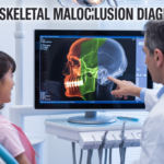
Our 3D TMJ (Temporomandibular Joint) reporting service offers advanced, high-resolution imaging analysis for accurate evaluation of joint structure and function. Using CBCT technology and expert radiological interpretation, we deliver detailed, precise, and clinically valuable reports to support confident diagnosis and treatment planning.
Why Choose Our 3D TMJ Reporting?
✔️ Advanced 3D Visualization Get a complete three-dimensional view of the TMJ, allowing better assessment of complex joint structures.
✔️ Expert Radiologist Review All reports are analyzed by experienced dental radiologists, ensuring accuracy and clinical reliability.
✔️ High Diagnostic Accuracy 3D imaging enables precise detection of joint abnormalities, bone changes, and structural variations.
✔️ Fast Turnaround Time Receive detailed reports quickly to support efficient workflow and timely patient care.
✔️ Safe & Precise Imaging We follow optimized CBCT protocols to ensure minimal radiation exposure with maximum clarity.
What We Analyze in 3D TMJ Reports
Condylar morphology and position
Joint space measurements
Cortical bone integrity
Degenerative changes and joint disorders
Asymmetry and alignment issues
Surrounding anatomical structures
Who Is It For?
Our 3D TMJ reporting services are ideal for:
General Dentists
Orthodontists
Oral & Maxillofacial Surgeons
Prosthodontists
Dental Clinics & Hospitals
Our Commitment
We combine cutting-edge 3D imaging technology, clinical expertise, and efficient reporting systems to deliver highly accurate and reliable TMJ assessments. Our goal is to enhance diagnostic confidence and improve patient outcomes through precise 3D TMJ reporting.